When epikeratoplasty is performed in an aphakic patient, it is termed epikeratophakia. This is a historical refractive procedure in which the corneal epithelium is removed and a lathed donor lenticule is placed onto the recipient cornea and secured with sutures until it heals into place. It has been referred to as "a living contact lens." It is no longer routinely performed given the advantages of modern laser refractive procedures.
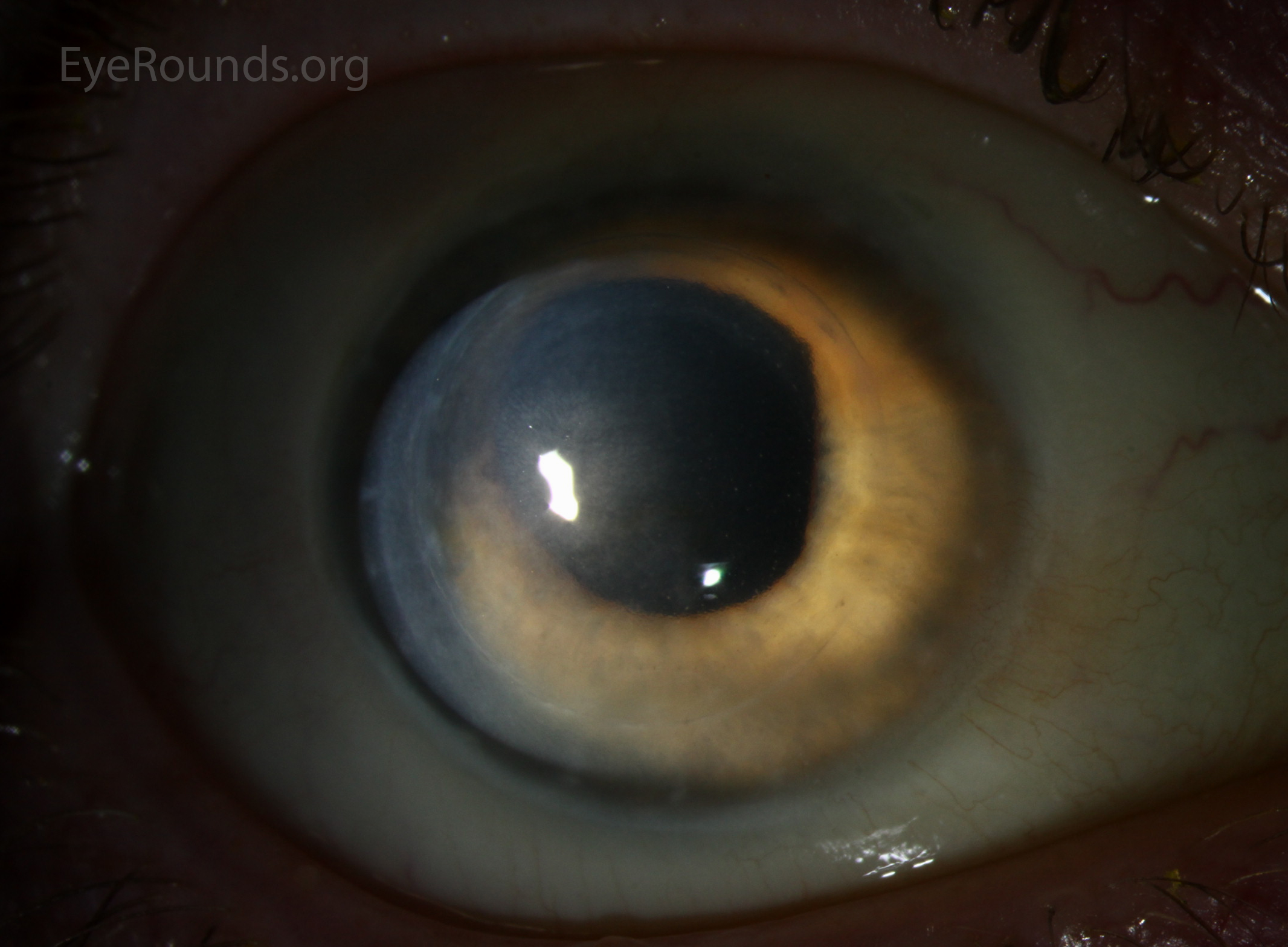
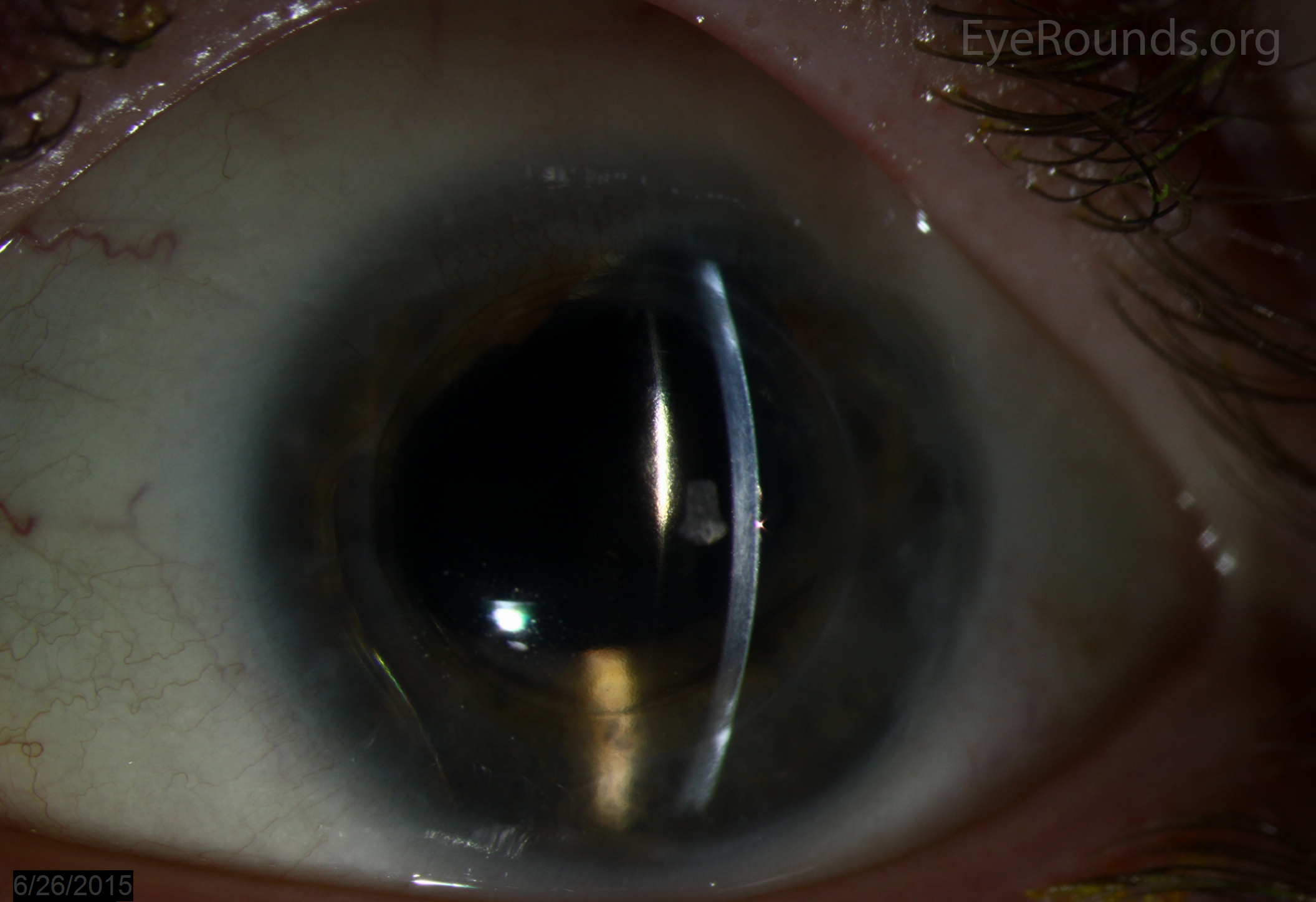
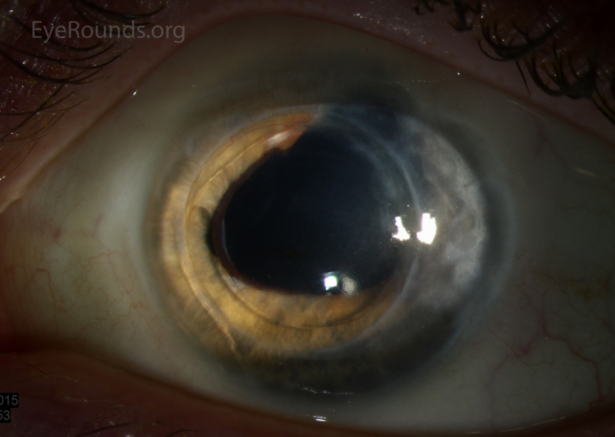
This patient is aphakic bilaterally secondary to removal of congenital cataracts as a child. He underwent the epikeratophakia procedure in both eyes 30 years prior to these photographs. The graft in the left eye was later removed and an anterior chamber intraocular lens was placed, though the original corneal scars from the epikeratoplasty are still visible in that eye. The right eye still has an epikeratophakia graft (easily visible on the anterior cornea in the slit beam photograph), but the vision is now limited by irregular astigmatism and corneal edema secondary to endothelial cell loss.

Ophthalmic Atlas Images by EyeRounds.org, The University of Iowa are licensed under a Creative Commons Attribution-NonCommercial-NoDerivs 3.0 Unported License.

Address
University of IowaLegal
Related Links