Expanded text by Brendan K. Penaluna, May 5, 2017
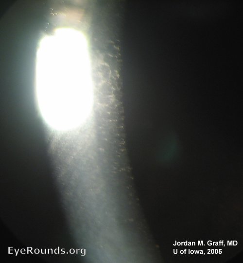
Fuchs' dystrophy is an example of a posterior corneal dystrophy. Thickening of the Descemet's membrane and defects of endothelial cell number and shape may result in progressive obscuration of vision. Cataract surgery in the setting of Fuch's dystrophy may rarely result in the decompensation of the corneal endothelium, creating an opportunity for the worsening of Fuchs' dystrophy. In this image of a patient with Fuchs' dystrophy post-cataract surgery, endothelial degeneration and subsequent edema and scarring can be appreciated as the white, hazy opacity contrasting against the brown iris and black of the pupil.[1]
Contributor: Jordan M. Graff, MD

Contributor: Andrew Doan, MD, PhD

Ophthalmic Atlas Images by EyeRounds.org, The University of Iowa are licensed under a Creative Commons Attribution-NonCommercial-NoDerivs 3.0 Unported License.