

Fuchs' epithelial-endothelial dystrophy of the cornea
Contributor: William Charles Caccamise, Sr, MD, Retired Clinical Assistant Professor of Ophthalmology, University of Rochester School of Medicine and Dentistry
*Dr. Caccamise has very generously shared his images of patients taken while operating during the "eye season" in rural India as well as those from his private practice during the 1960's and 1970's. Many of his images are significant for their historical perspective and for techniques and conditions seen in settings in undeveloped areas.
Category: Cornea
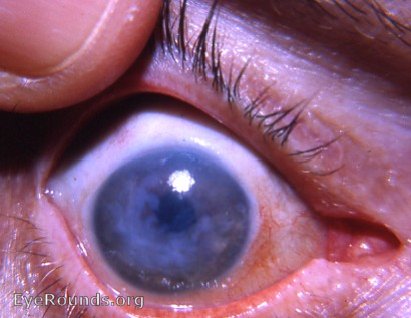
The cornea of this aphakic eye demonstrates decompensation typical of that with Fuchs' epithelial-endothelial dystrophy.

Fuchs epithelial-endothelial corneal dystrophy with central ruptured epithelial bullae
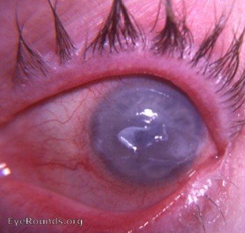
This genetically-based corneal dystrophy occurs more frequently in females in the 5th and 6th decades of life. It is initally manifested by a deficiency in the corneal endothelium which is first detected on slit-lamp examination by specular reflection. Later, the epithelium participates in the dystrophic process. Eventually, the corneal pathology may reach the advanced stage evident in the photo. Here, central epithelial bullae have broken down.

Ophthalmic Atlas Images by EyeRounds.org, The University of Iowa are licensed under a Creative Commons Attribution-NonCommercial-NoDerivs 3.0 Unported License.