

Groenouw's Type I Granular corneal dystrophy
Contributor: William Charles Caccamise, Sr, MD, Retired Clinical Professor of Ophthalmology, University of Rochester School of Medicine and Dentistry
*Dr. Caccamise has very generously shared his images of patients taken while operating during the "eye season" in rural India as well as those from his private practice during the 1960's and 1970's. Many of his images are significant for their historical perspective and for techniques and conditions seen in settings in undeveloped areas.
Category: Cornea
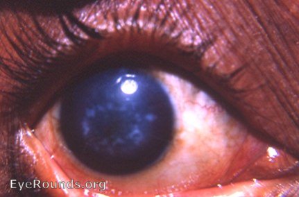
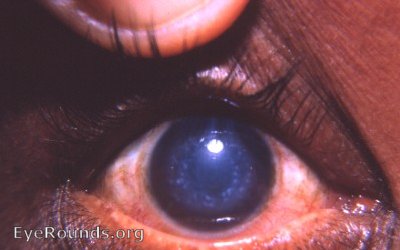
From a clinical point of view, granular corneal dystrophy can be distinguished from macular corneal dystrophy by the slit-lamp findings: the opcites of the former are sharp and the adjacent stroma is clear without scarring.In the macular form, the opacities have fuzzy edges.
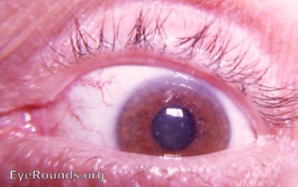
 This corneal dystrophy occurs in the patient, his father, and his brother.
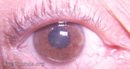
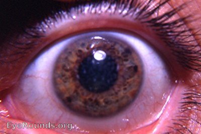
An unrelated case

Ophthalmic Atlas Images by EyeRounds.org, The University of Iowa are licensed under a Creative Commons Attribution-NonCommercial-NoDerivs 3.0 Unported License.