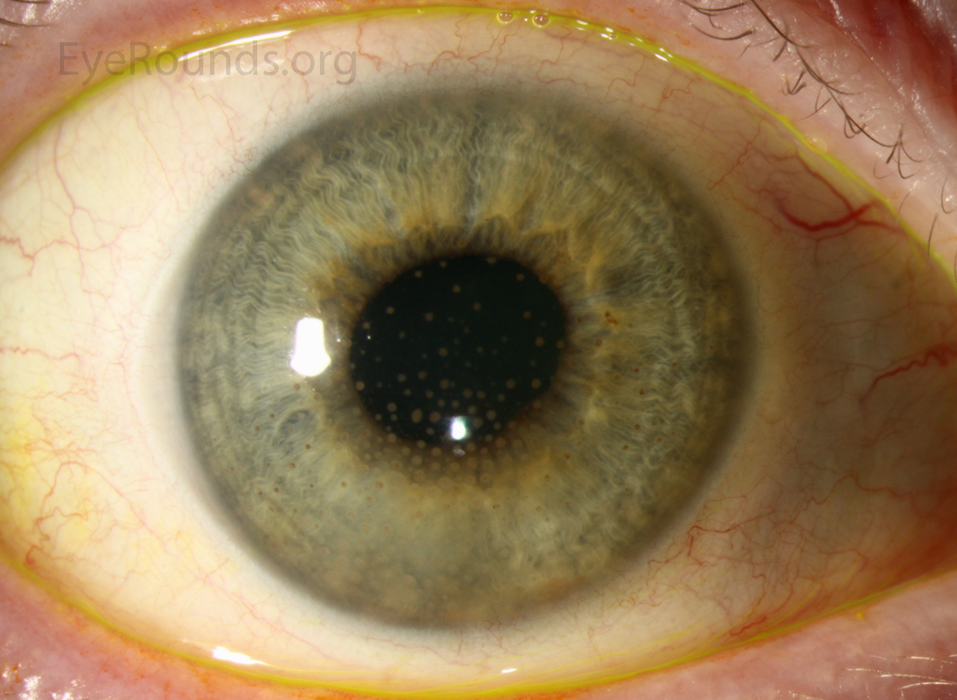
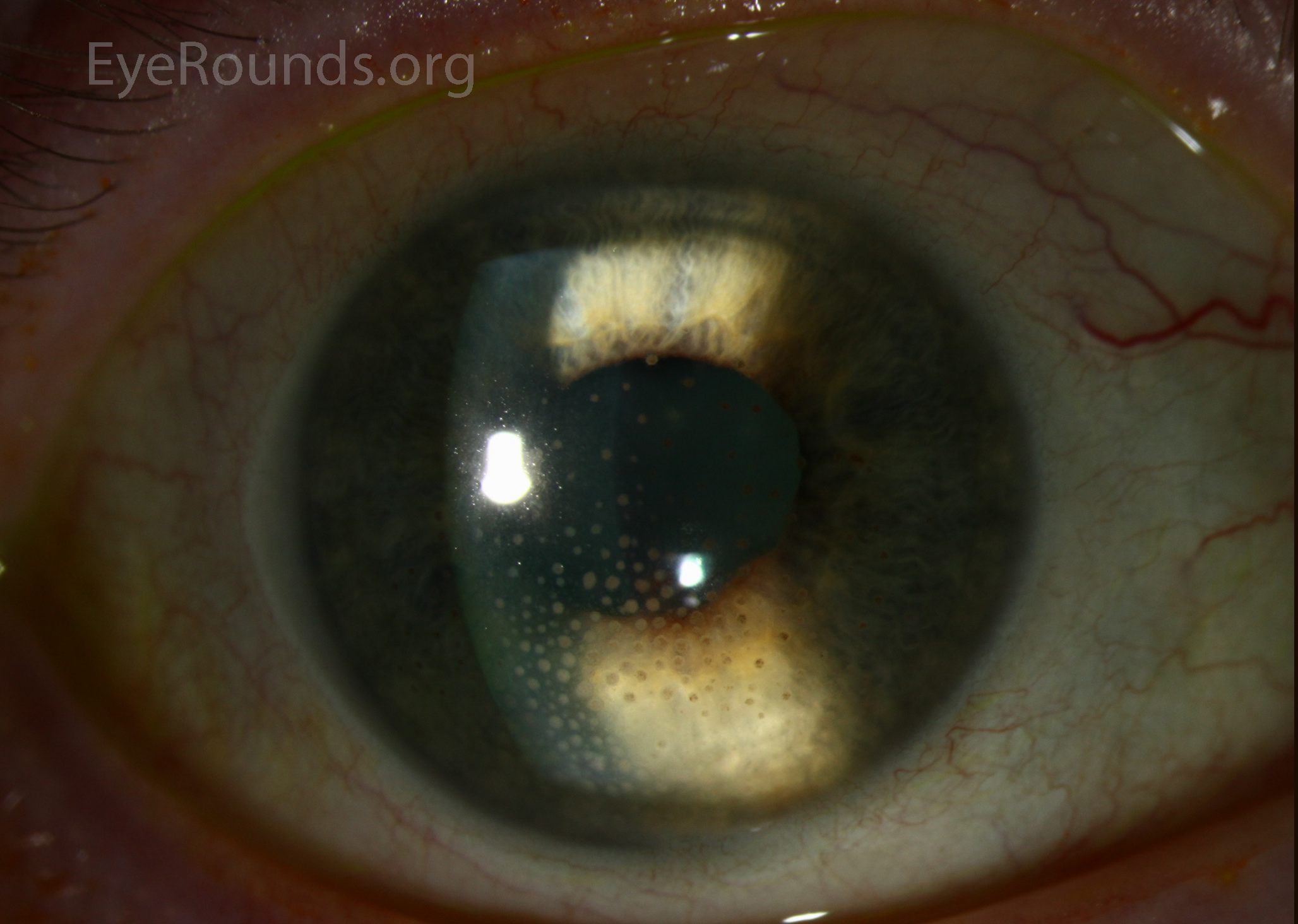
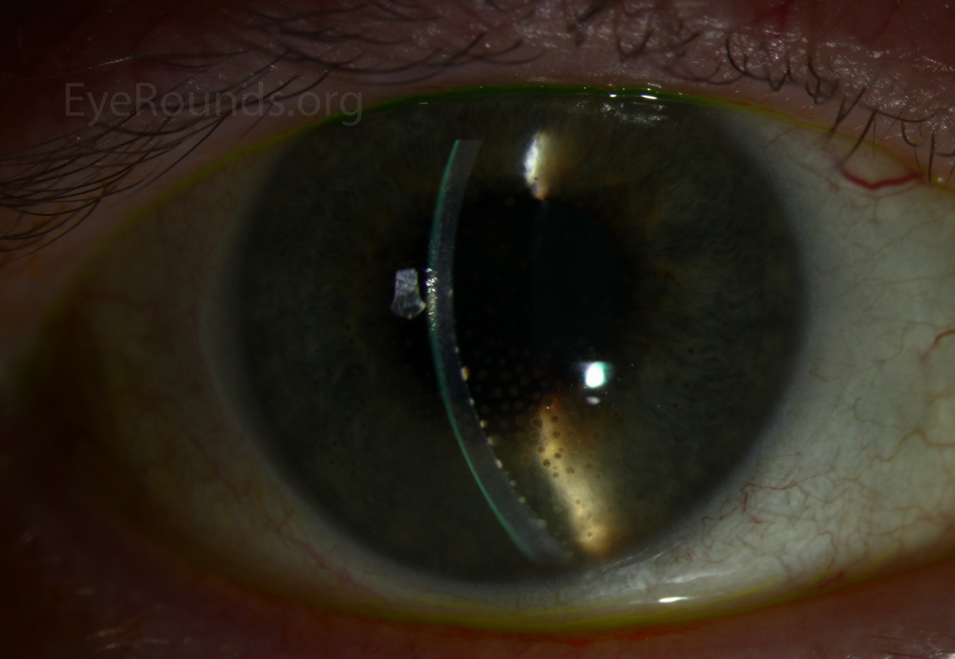
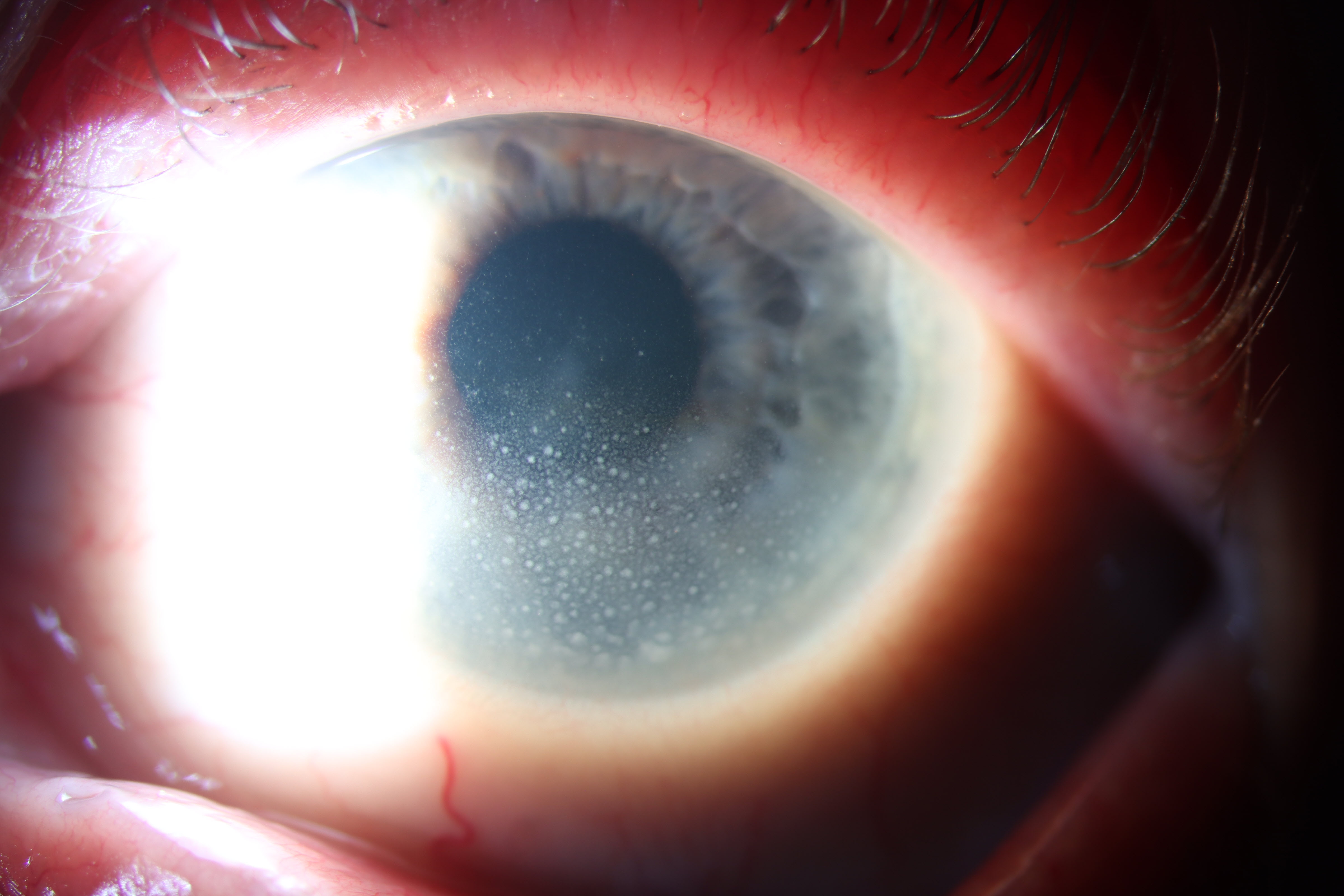
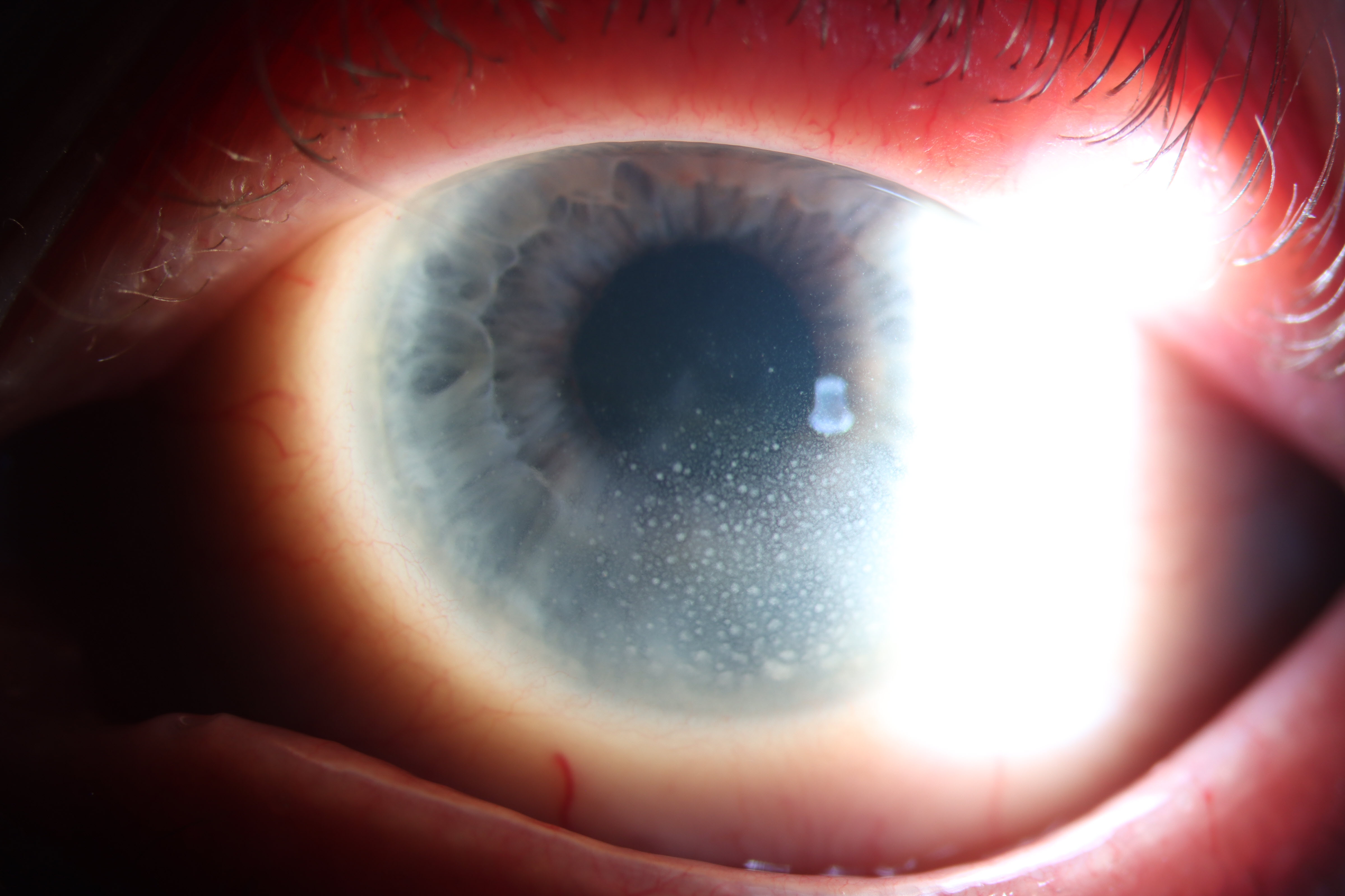
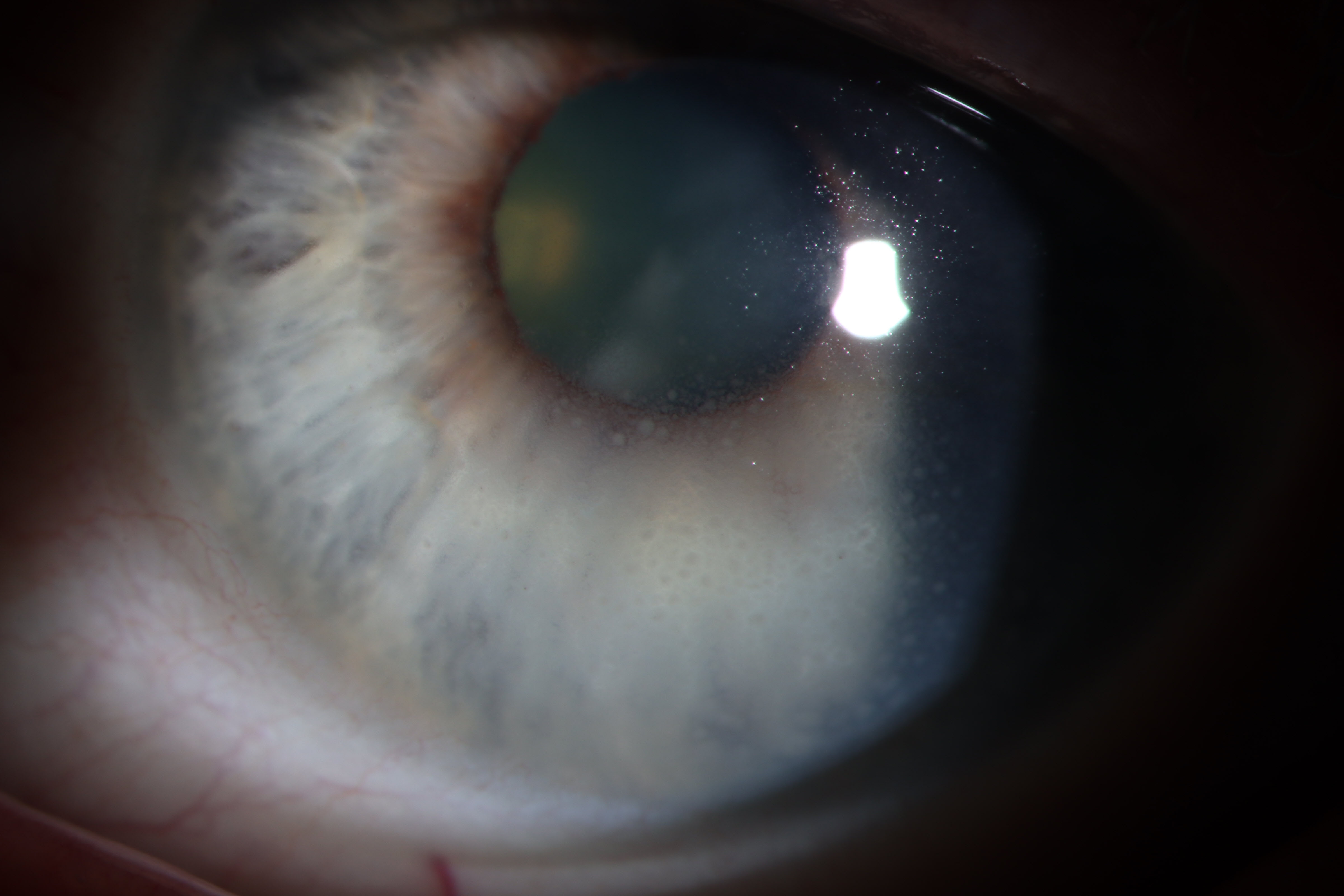
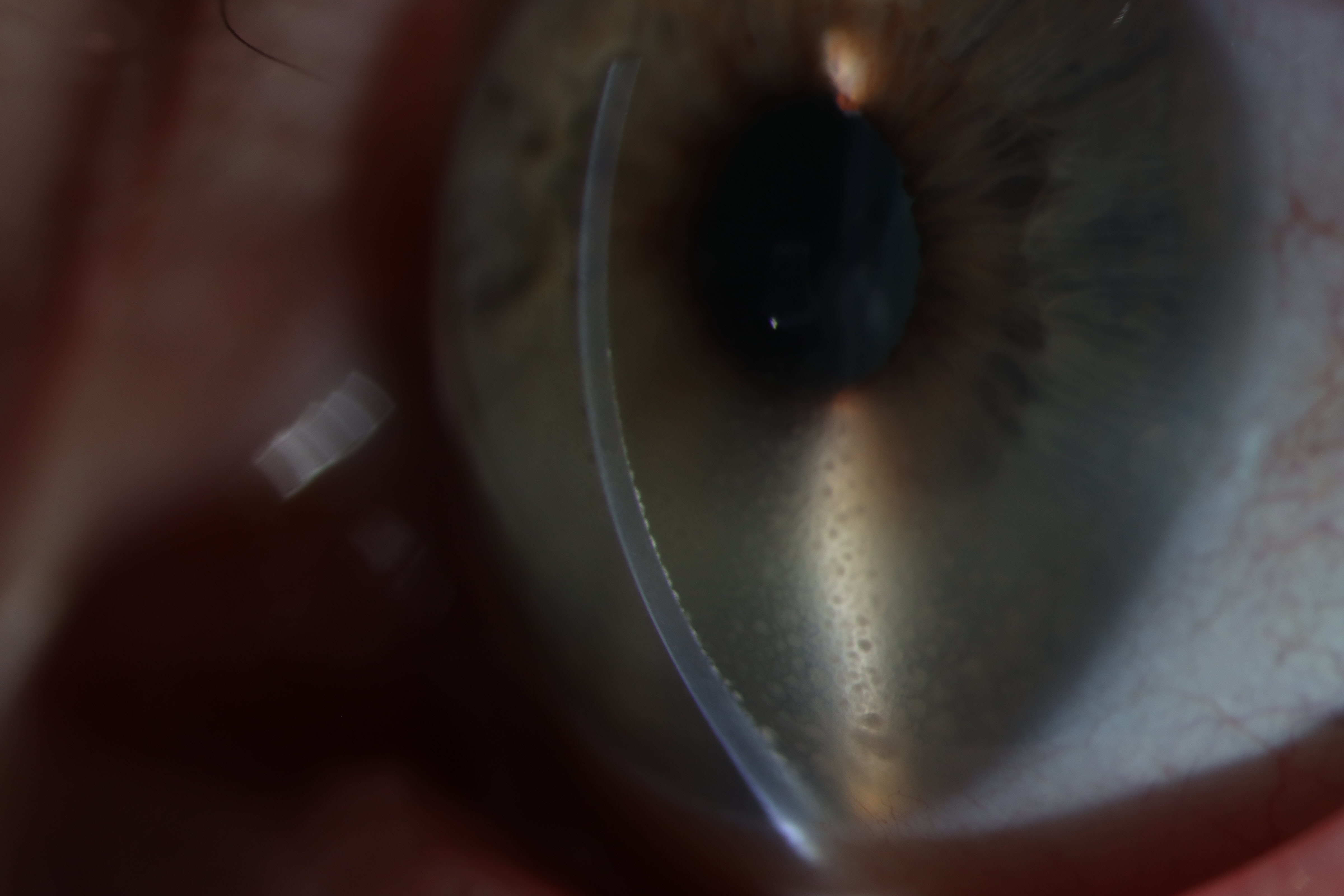
This is a 51-year-old female that presented with severe ocular pain and photophobia. She presented acutely and had evidence of mutton-fat keratic precipitates (KP) on exam. She was later diagnosed with sarcoidosis. KP are collections of inflammatory cells on the corneal endothelium. When these coalesce and become large with a yellowish color, they are described as mutton-fat KP. These are typically associated with granulomatous inflammation.
A 68-year-old female presented to the emergency department with a chief complaint of blurry vision in the left eye. Slit lamp examination was significant for 2+ cell, flare, and mutton fat keratic precipitates in the distribution of Arlt’s triangle. The posterior segment exam was unremarkable. Workup was significant for positive HSV2 serum IgG. Keratic precipitates are clumps of inflammatory cells, that due to gravity and convection currents within the anterior chamber, settle in a wedge-shaped distribution on the inferior corneal endothelium. They are typically associated with, although not pathognomonic for, granulomatous inflammation.

Ophthalmic Atlas Images by EyeRounds.org, The University of Iowa are licensed under a Creative Commons Attribution-NonCommercial-NoDerivs 3.0 Unported License.

Address
University of IowaLegal
Related Links