
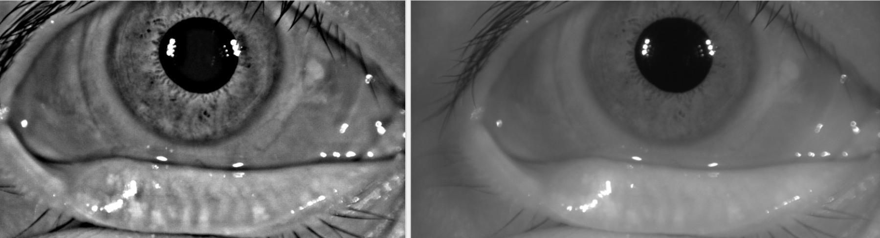
Often invisible under the palpebral conjunctiva, the meibomian glands make the oil layer of the tear film. In meibomian gland disease, the glands often widen in response to stress and then begin to atrophy.
Figures 2 through 4. Contributor & Photographer: Tressa Larson OD FAAO
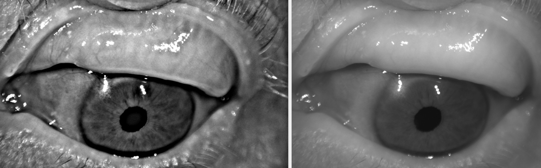
In meibomian gland disease, the glands often widen in response to stress and then begin to atrophy. Infrared images such the ones in figure 2 allow better visualization of the meibomian glands than the white light view. The right is the original image and the left is an enhanced version. This patient has severe meibomian gland disease. The glands nasal and temporal have dropped out entirely and the remaining glands are very short and tortuous.

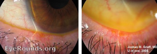
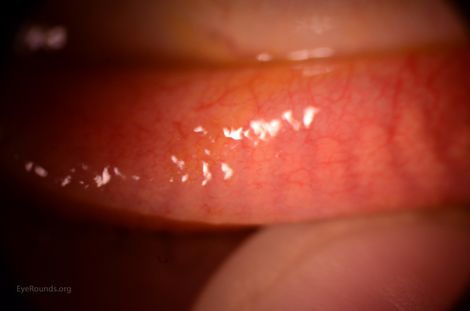
In these images it is possible to see the blunted meibomian glands on the lower lid and to see that they only extend about a quarter or halfway up the tarsal plate.

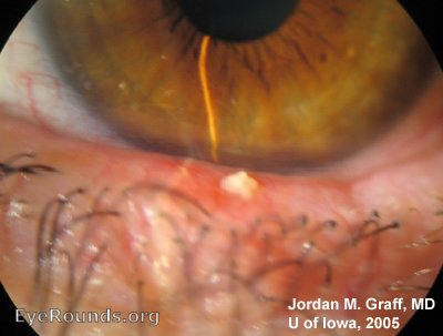
This patient had such severe meibomian gland disease that the glands are completely atrophied. You can see where the glands used to be.

Ophthalmic Atlas Images by EyeRounds.org, The University of Iowa are licensed under a Creative Commons Attribution-NonCommercial-NoDerivs 3.0 Unported License.

Address
University of IowaLegal
Related Links

