EyeRounds Online Atlas of Ophthalmology
Contributor: William Charles Caccamise, Sr, MD, Retired Clinical Assistant Professor of Ophthalmology, University of Rochester School of Medicine and Dentistry
*Dr. Caccamise has very generously shared his images of patients taken while operating during the "eye season" in rural India as well as those from his private practice during the 1960's and 1970's. Many of his images are significant for their historical perspective and for techniques and conditions seen in settings in undeveloped areas.
Category: Cataract
Morgagnian hypermature cataract: a rare shrinking stage

Very rarely a Morgagnian hypermature cataract will follow the course that the cataract in the photo has chosen: a mature corticonuclear cataract will become a Morgagnian hypermature cataract with all of its cortex liquefied; the posterior capsule will develop leakage that permits the liquefied cortex to leave the lens gradually; finally so much of the liquefied cortex has leaked out that only the lens capsule and the encapsulated nucleus remain. In the photo, the subluxated, encapsulated nucleus is seen. The scattered subcapsular white dots indicate that there is still some retained cortex. Notice the dark space between the superior border of the shrunken lens and the pupil margin superiorly - an indication of subluxation of the abnormal lens. In 1966 when this photo was taken, the operation of choice was a Snellen maneuver with a lens loop and with a hope that no liquified vitreous would be lost.

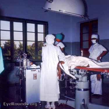
One of the 4 ORs available for eye cases at the Kurji Holy Family Hospital Hospital Eye Clinic

Ophthalmic Atlas Images by EyeRounds.org, The University of Iowa are licensed under a Creative Commons Attribution-NonCommercial-NoDerivs 3.0 Unported License.


