This site uses tracking information. Visit our privacy policy. Click to agree to this policy and not see this again.

Ophthalmology and Visual Sciences

Neovascularization of the iris (NVI) secondary to a central retinal artery occlusion (CRAO)

Neovascularization of the iris (NVI) secondary to a central retinal artery occlusion (CRAO)

Category(ies): Glaucoma/ Iris and Retina
Contributor: Andrew Goldstein, MD; James C. Folk, MD
Photographer: Sarah Skiles, CRA

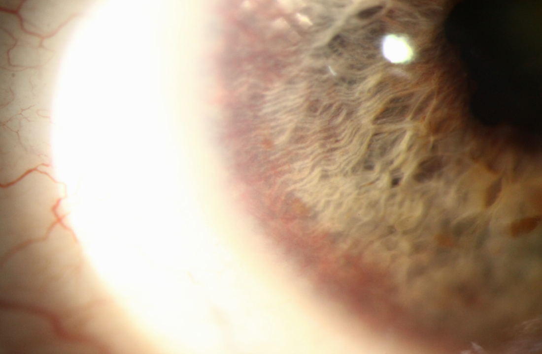
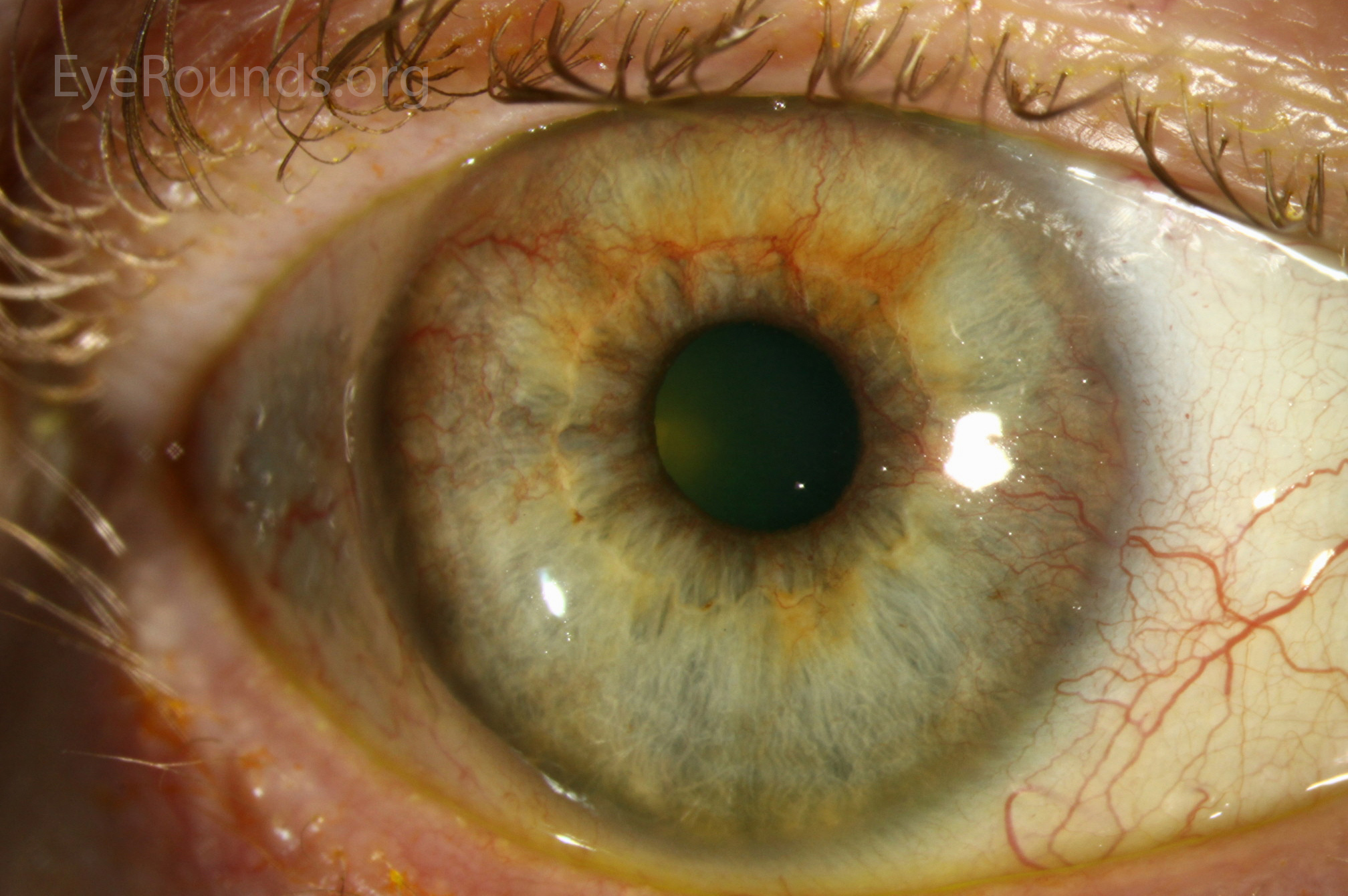
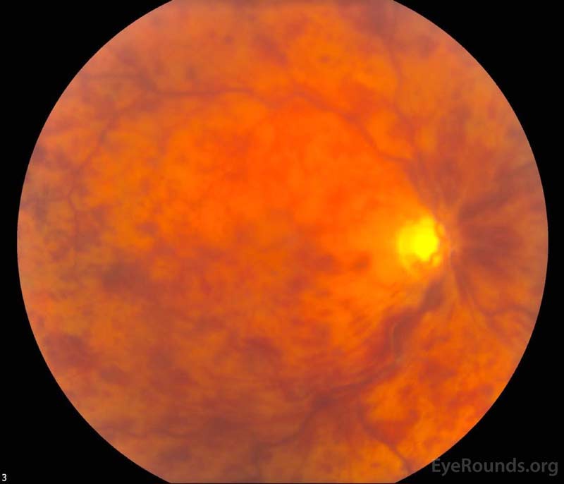
This patient initially presented with neovascularization of the iris (NVI), or rubeosis iridis, and neovascular glaucoma (NVG) that occurred three months after a central retinal artery occlusion (CRAO). The appearance of the NVI in this patient is subtle as he has undergone intravitreal injections of bevacizumab for neovascular glaucoma. Although much less common than in central retinal vein occlusions, the incidence of NVI has been shown to be about 10.9% after a CRAO.

diffuse illumination of the right eye shows NVI as demonstrated by the fine, lacy blood vessels surrounding the pupillary margin
Figure 1: Anterior segment color photography with diffuse illumination of the right eye shows NVI as demonstrated by the fine, lacy blood vessels surrounding the pupillary margin. A slightly enhanced view of the NVI is shown within the white circle.
alt text
Figure 2: Anterior segment color photography with direct illumination and higher magnification of the pupillary margin shows the fine blood vessels that are characteristic of NVI (peripheral edge marked by white arrows).

Category(ies): Glaucoma/ Iris and Retina
Contributor: Patrick Donegan MD, Elaine Binkley MD
Photographer: Nicole Radunzel, CRA

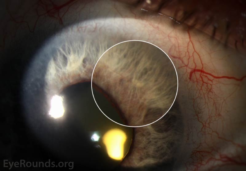
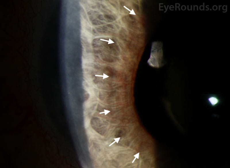
This is a 65-year-old woman with history of central retinal artery occlusion one month prior who presented with neovascular glaucoma. As displayed in the slit lamp images, she had dense inferior neovascularization of the angle accompanying her near 360-degree neovascularization of the iris.

diffuse illumination of the right eye shows NVI as demonstrated by the fine, lacy blood vessels surrounding the pupillary margin
Figure 1: Anterior segment color photography with diffuse illumination of the right eye shows NVI as demonstrated by the fine, lacy blood vessels surrounding the pupillary margin. A slightly enhanced view of the NVI is shown within the white circle.
alt text
Figure 2: Anterior segment color photography with direct illumination and higher magnification of the pupillary margin shows the fine blood vessels that are characteristic of NVI (peripheral edge marked by white arrows).
Reference:

Jung YH, Ahn SJ, Hong JH, et al. Incidence and Clinical Features of Neovascularization of the Iris following Acute Central Retinal Artery Occlusion. Korean J Ophthalmol. 2016;30(5):352-359. doi:10.3341/kjo.2016.30.5.352


Image Permissions:
Creative Commons

Ophthalmic Atlas Images by EyeRounds.org, The University of Iowa are licensed under a Creative Commons Attribution-NonCommercial-NoDerivs 3.0 Unported License.