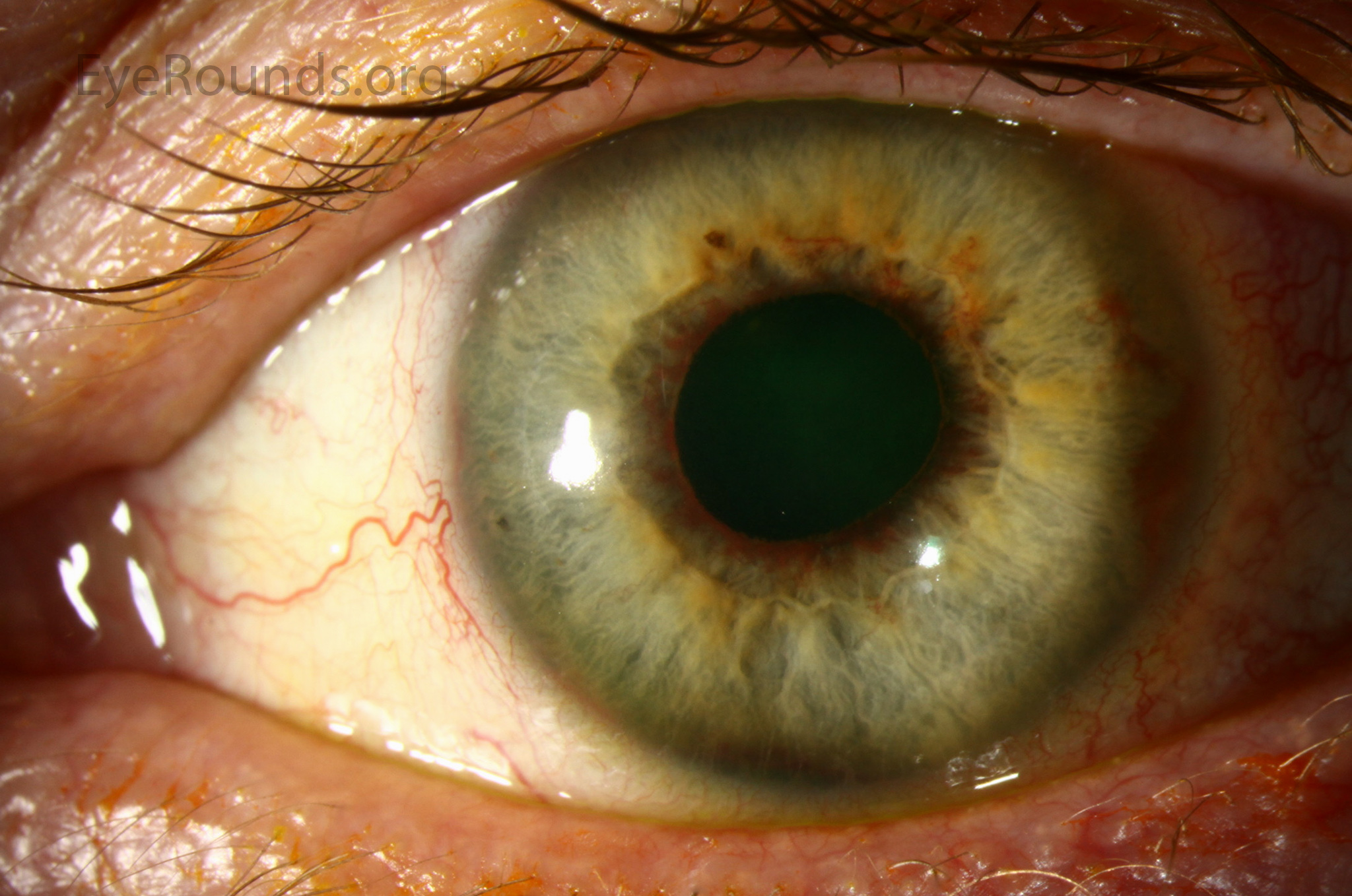
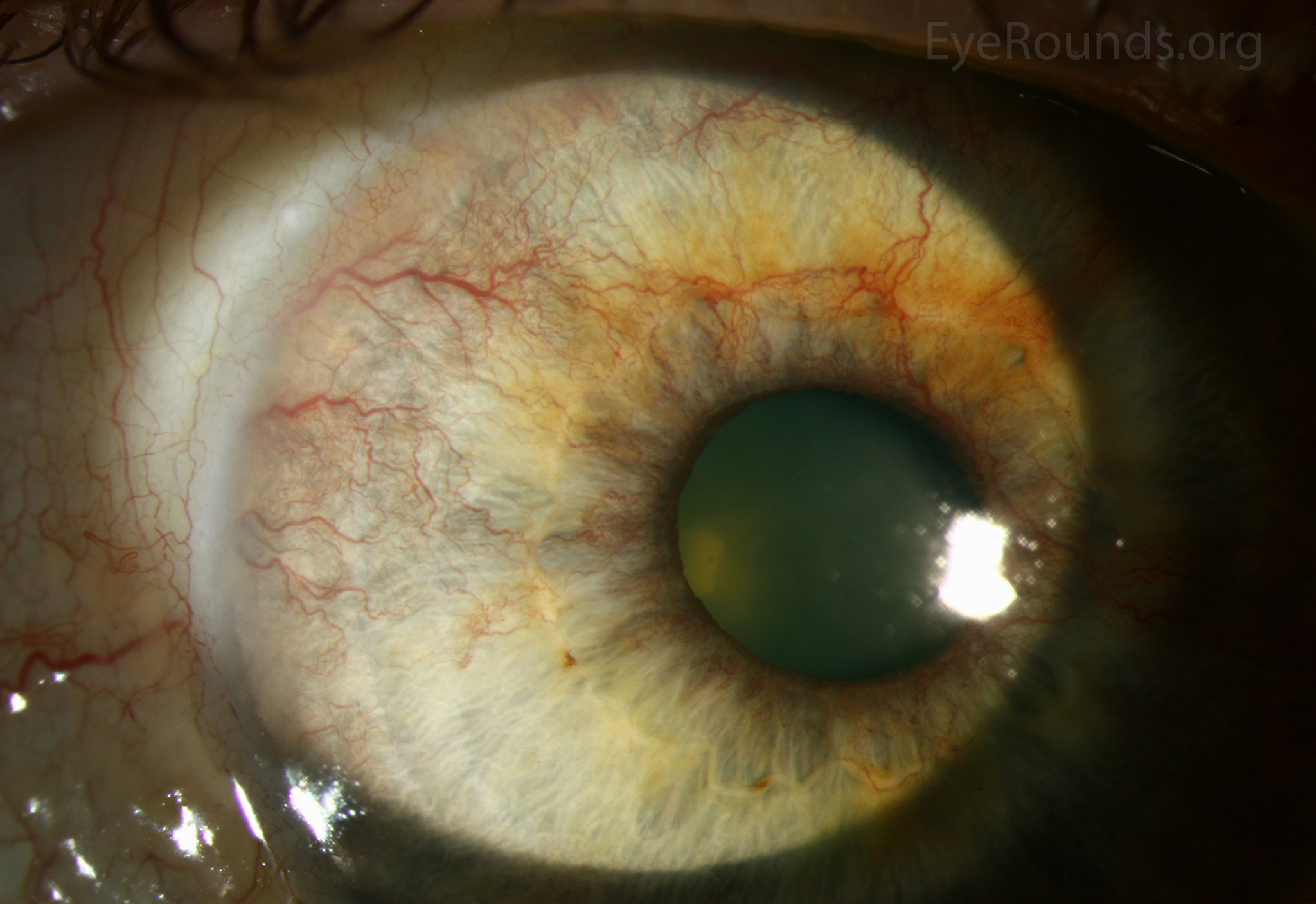
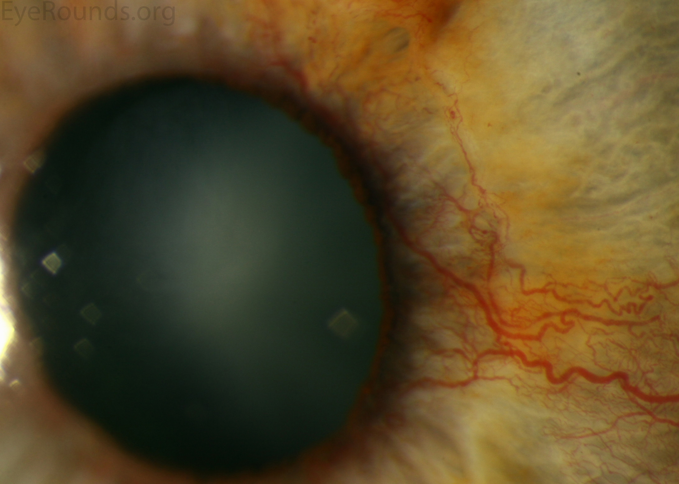
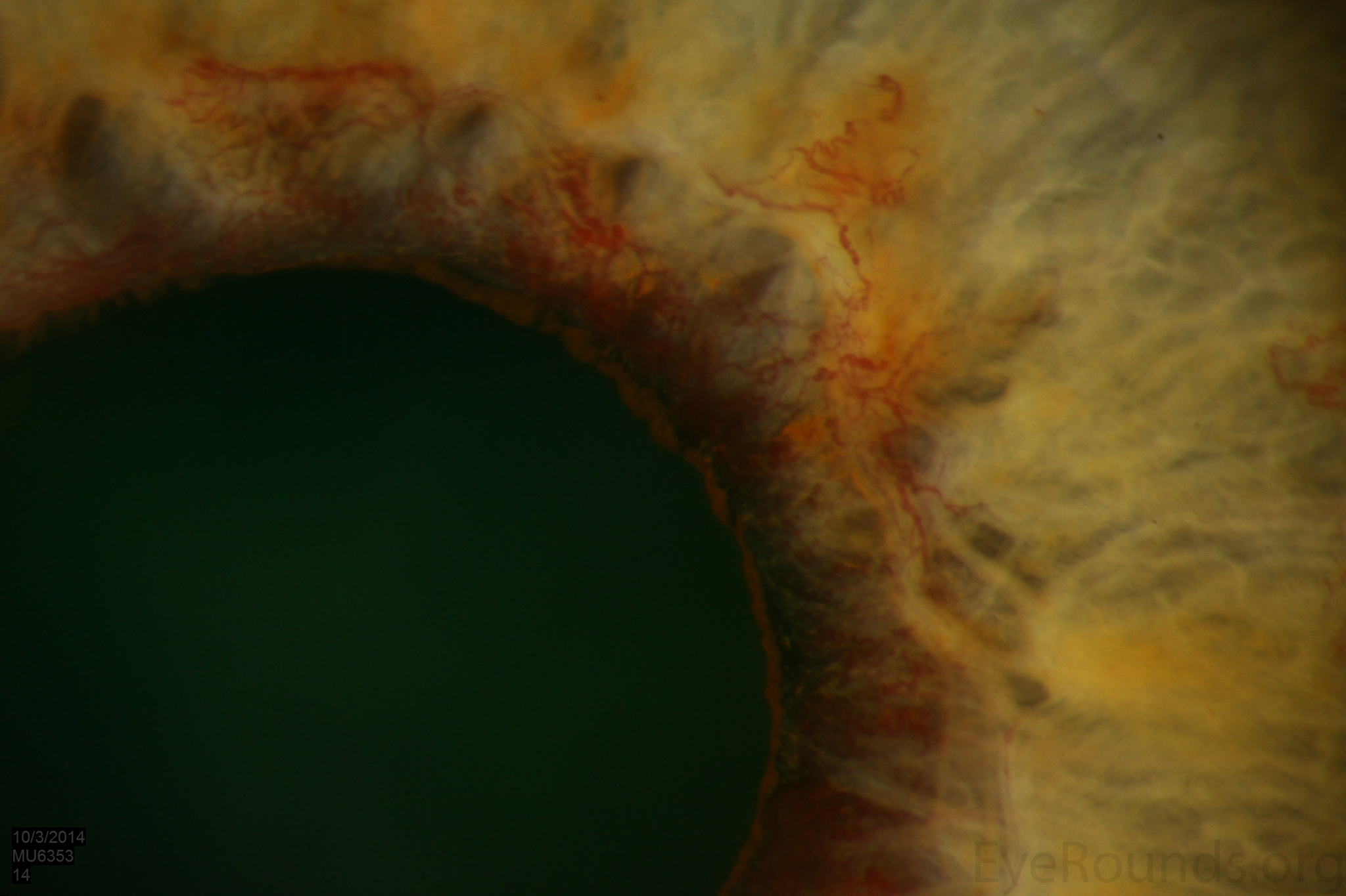
This is a 57-year-old man with a past medical history of diabetes mellitus type 2, who presented to the ophthalmology clinic for decreased vision. His most recent hemoglobin A1c was 12.8%. Upon applanation his intraocular pressures were 11 mmHg OD and 32 mmHg OS. Both eyes had fine vessels coursing along the iris surface in an irregular path directed radially towards the angle. His anterior segment exam demonstrated tufts of vessels along the pupillary margin in the left eye and fine vessel extending from the angle at 3 o'clock. There was a 1 mm layered hyphema in the left eye. On gonioscopy, he had a 35 degree angle and 360 degrees of neovascularization of the angle in both eyes. Posterior segment exam showed severe proliferative diabetic retinopathy in both eyes.
Neovascularization of the iris (NVI), also known as rubeosis iridis, is when small fine, blood vessels develop on the anterior surface of the iris in response to retinal ischemia. These changes most often develop at the pupillary border, but it is important to perform gonioscopy in order to investigate for involvement of the angle. Patients with NVI are prone to spontaneous hyphemas as these blood vessels are fragile and lend to bleeding. Patients with proliferative diabetic retinopathy who develop NVI are often treated with panretinal photocoagulation with or without an intravitreal injection of an anti-VEGF medication.

Ophthalmic Atlas Images by EyeRounds.org, The University of Iowa are licensed under a Creative Commons Attribution-NonCommercial-NoDerivs 3.0 Unported License.

Address
University of IowaLegal
Related Links