A rhegmatogenous retinal detachment results when liquified vitreous passes through a retinal break into the potential subretinal space.
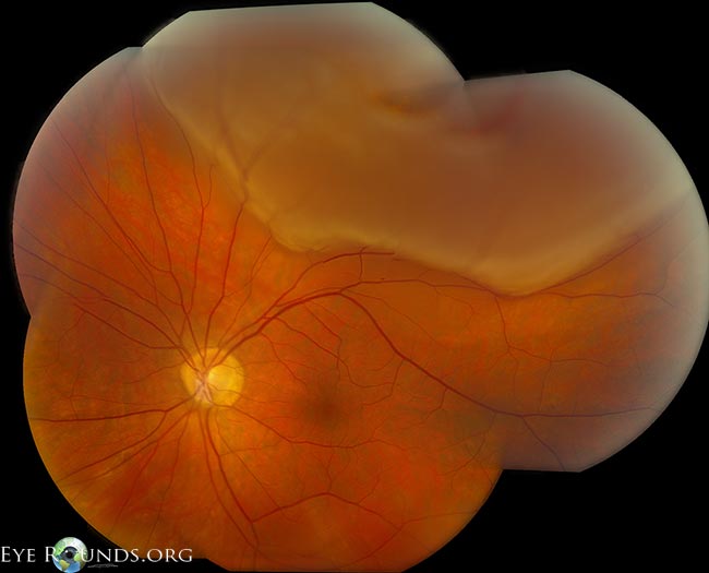
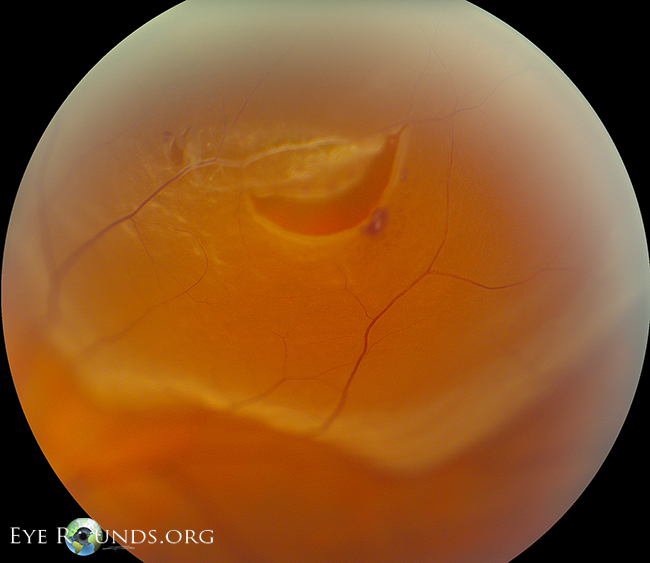
71-year-old man presented with 2 weeks of blurred vision and curtain-like vision loss obscuring the inferior portion of his left visual field. Visual acuity was 20/50+2. Fundoscopy (see image) demonstrated a large horseshoe break at 12:30 at the equator, with a slightly rolled edge and a bridging vessel. There was subretinal fluid from 10:00 clockwise to 3:00, extending posteriorly to just involve the center of the fovea. This macula-involving rhegmatogenous retinal detachment was repaired surgically. Three months later, his vision improved to 20/20.


This is a patient who presented with an inferior visual field defect and multiple flashes and floaters. These images demonstrate an excellent example of a large, superior horseshoe tear with a bullous retinal detachment.
These OCT images show an inferior, macula-off, chronic rhegmatogenous retinal detachment.

Ophthalmic Atlas Images by EyeRounds.org, The University of Iowa are licensed under a Creative Commons Attribution-NonCommercial-NoDerivs 3.0 Unported License.