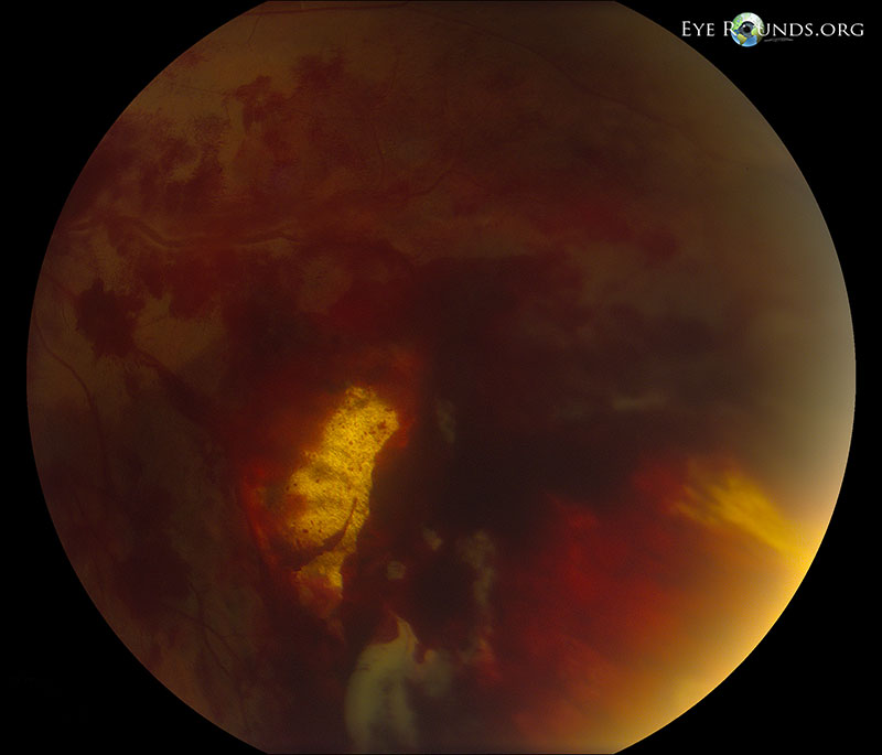
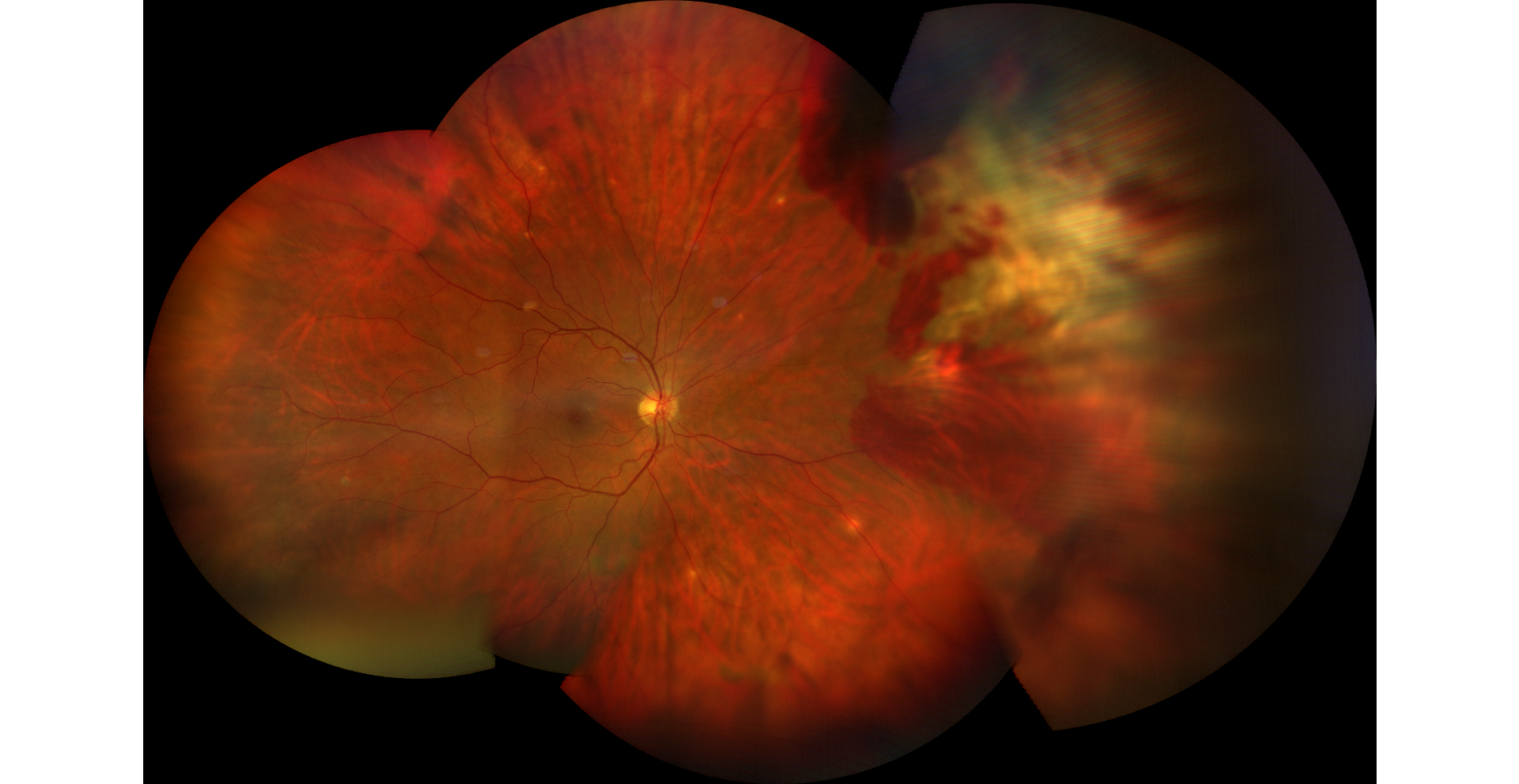
18-year-old male who suffered an ocular injury from a BB discharged from a BB gun. The BB entered the medial aspect of the orbit, but did not penetrate the globe. The photo shows inferonasal sclopetaria with associated intraretinal and pre-retinal hemorrhage. Sclopetaria is often seen after high-velocity injury to the orbit. Also note the extensive commotio retinae, or retinal whitening, especially in the macula and around the optic disc. No retinal holes or tears were identified.
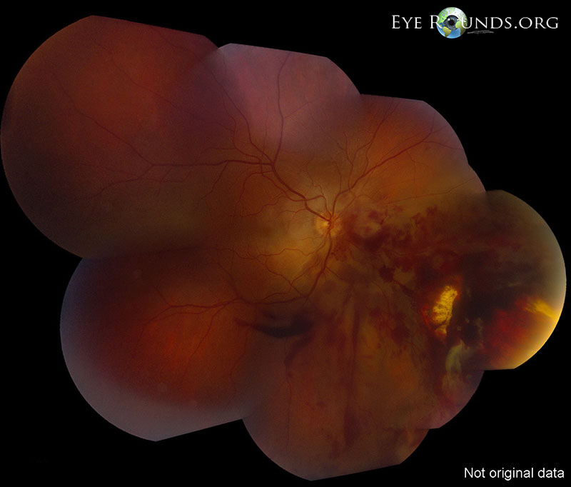
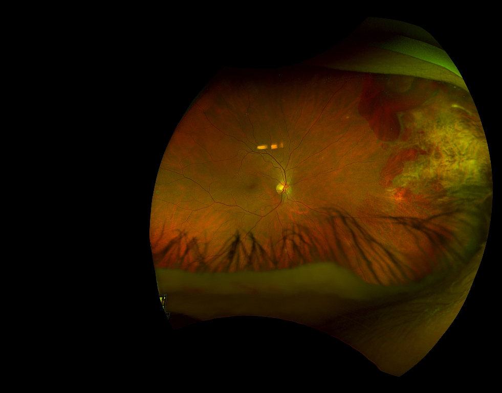
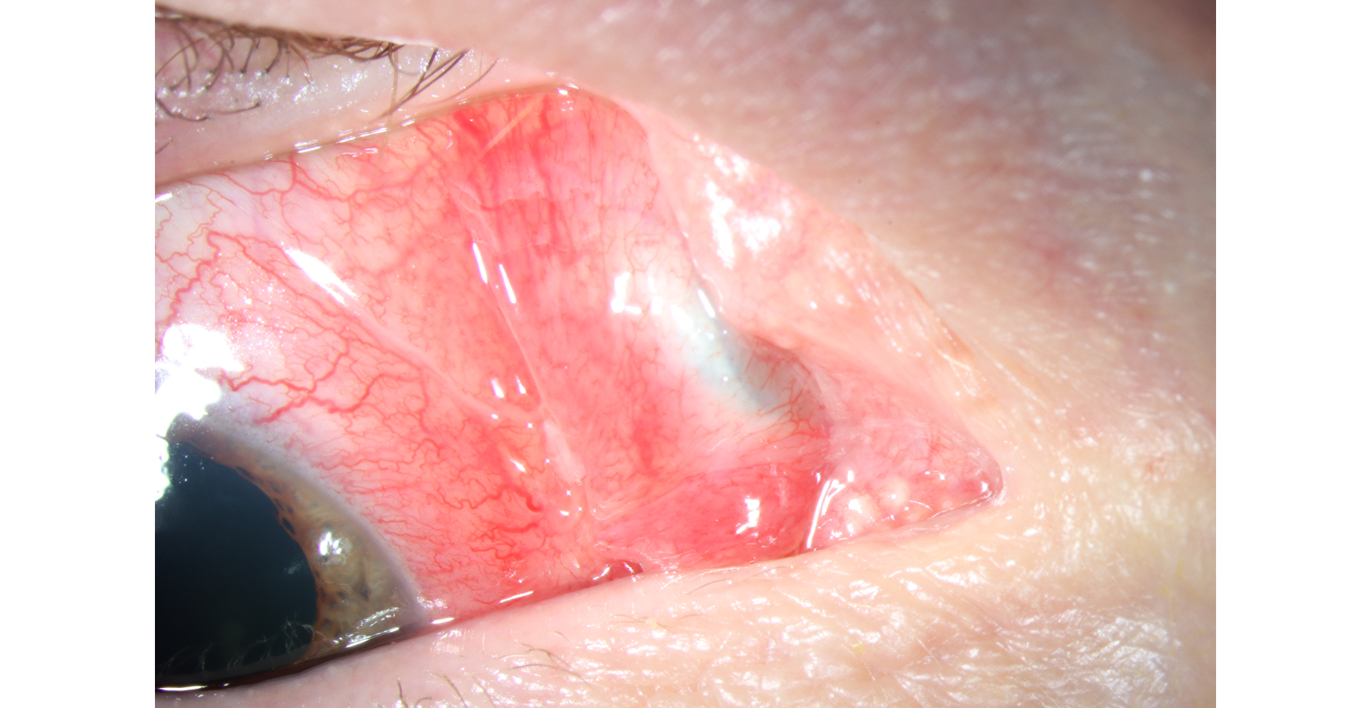
A woman in her 40s presented due to persistent redness and foreign body sensation several weeks after an ocular injury from a pellet gun. She was found to have a nasal subconjunctival metallic foreign body with scarring of the plica semilunaris (Figure 1). Dilated fundus exam showed mild vitreous hemorrhage and a nasal peripheral region of hypopigmented scarring as well as intra- and subretinal hemorrhage, consistent with chorioretinitis sclopetaria (Fgiure 2). Chorioretinitis sclopetaria is a full-thickness rupture and retraction of the retina and choroid due to blunt trauma from a high velocity projectile, often leaving visible bare sclera at the injury site. This patient’s subconjunctival pellet was successfully surgically removed, and there was no evidence of scleral laceration or intraocular penetration. Ultra-widefield imaging on follow up 2 weeks later showed interval improvement in the intra- and subretinal hemorrhage with chorioretinal atrophy (Figure 3).

Ophthalmic Atlas Images by EyeRounds.org, The University of Iowa are licensed under a Creative Commons Attribution-NonCommercial-NoDerivs 3.0 Unported License.


Address
University of IowaLegal
Related Links