EyeRounds Online Atlas of Ophthalmology
Contributor: William Charles Caccamise, Sr, MD, Retired Clinical Assistant Professor of Ophthalmology, University of Rochester School of Medicine and Dentistry
*Dr. Caccamise has very generously shared his images of patients taken while operating during the "eye season" in rural India as well as those from his private practice during the 1960's and 1970's. Many of his images are significant for their historical perspective and for techniques and conditions seen in settings in undeveloped areas.
Category: External Disease
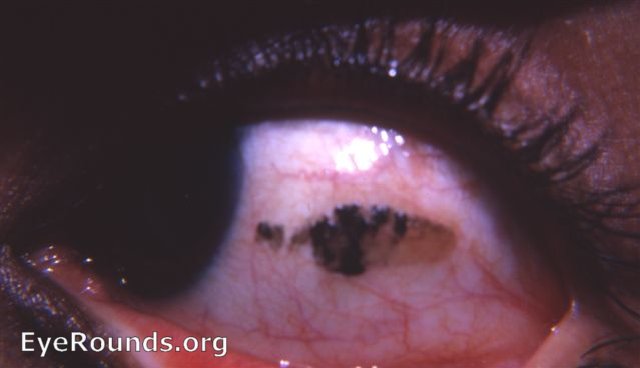
Blackened Bitot's spot in Vitamin A deficiency

The classic Bitot's spot consists of whipped up meibomian secretions and conjunctival debris with bacteria that adhere to a xerotic patch of conjunctiva. It is usually but not always located temporally.It can be removed with a Q-tip to reveal the underlying xerosis. It quickly reforms.The clinician in India automatically suspects a diagnosis of Vitamin A deficiency when the patient presents with Bitot's spots - they are usually bilateral. However, Dr. Caccamise did have a patient in Rochester, NY with a subtle Bitot's spot due to a patch of conjunctival xerosis and no Vitamin A deficiency.
Why is this Bitot's spot black? In India, kohl - a powder of antimony or other black pigment in mascara form - is a preparation used as a cosmetic around the eyes - even in infants. My rural patients often stated that it was used to ward off the evil eye. This is blackened type of Bitot's spot that has rarely if ever been mentioned in an atlas.


Ophthalmic Atlas Images by EyeRounds.org, The University of Iowa are licensed under a Creative Commons Attribution-NonCommercial-NoDerivs 3.0 Unported License.


