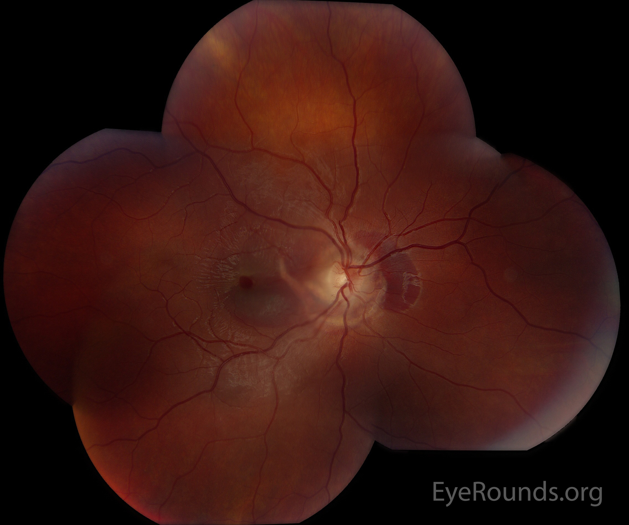
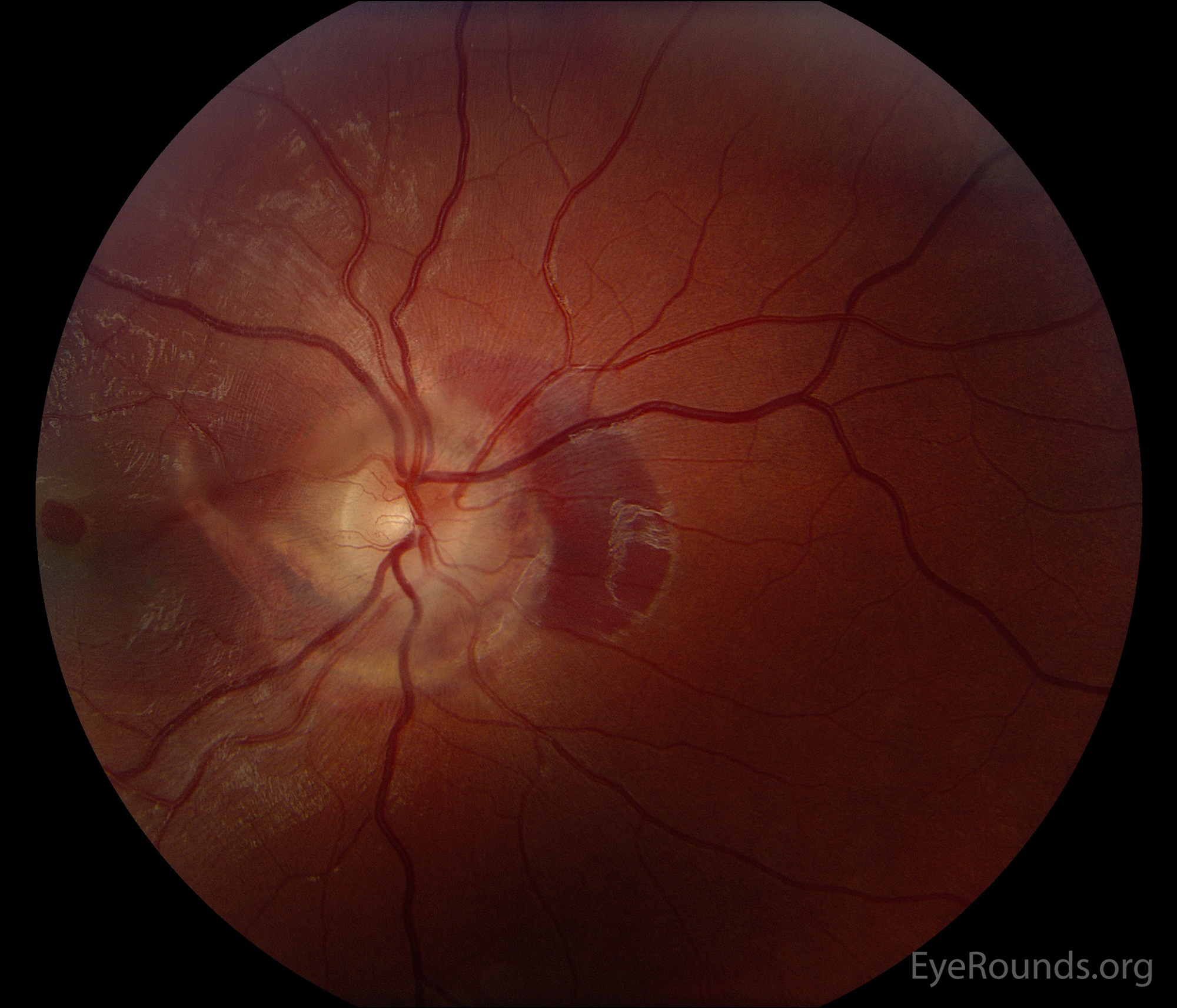
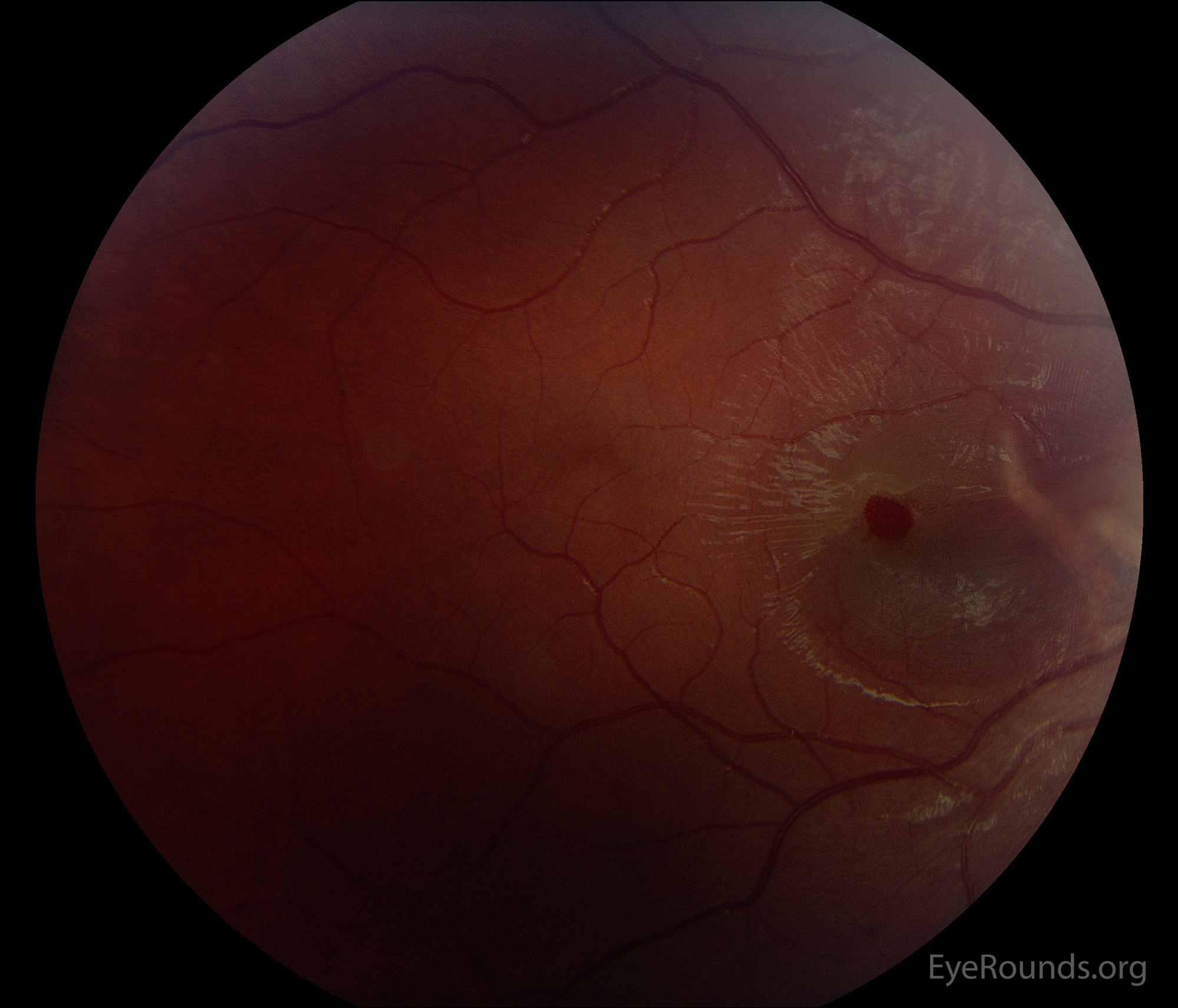
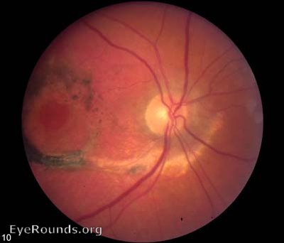
This patient experienced blunt eye injury after bungee cord recoil. Color fundus montage photograph of the right eye shows a macular hole and peripapillary choroidal ruptures extending into the macula. There is subretinal hemorrhage and fluid inferior to the macular hole and in the peripapillary region. Optical coherence tomography (Heidelberg) demonstrates a partial-thickness macular hole with subretinal heme.
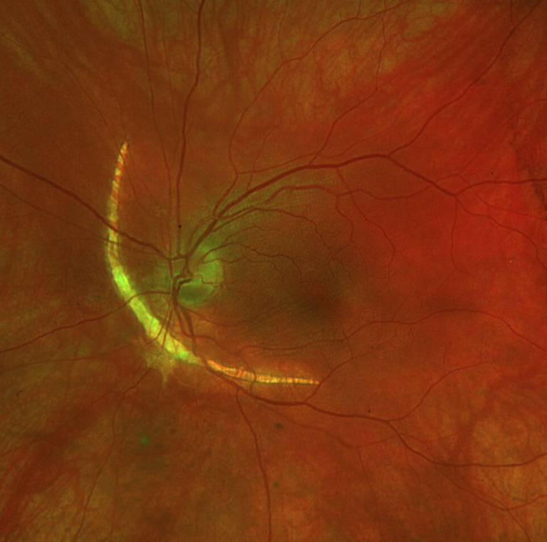
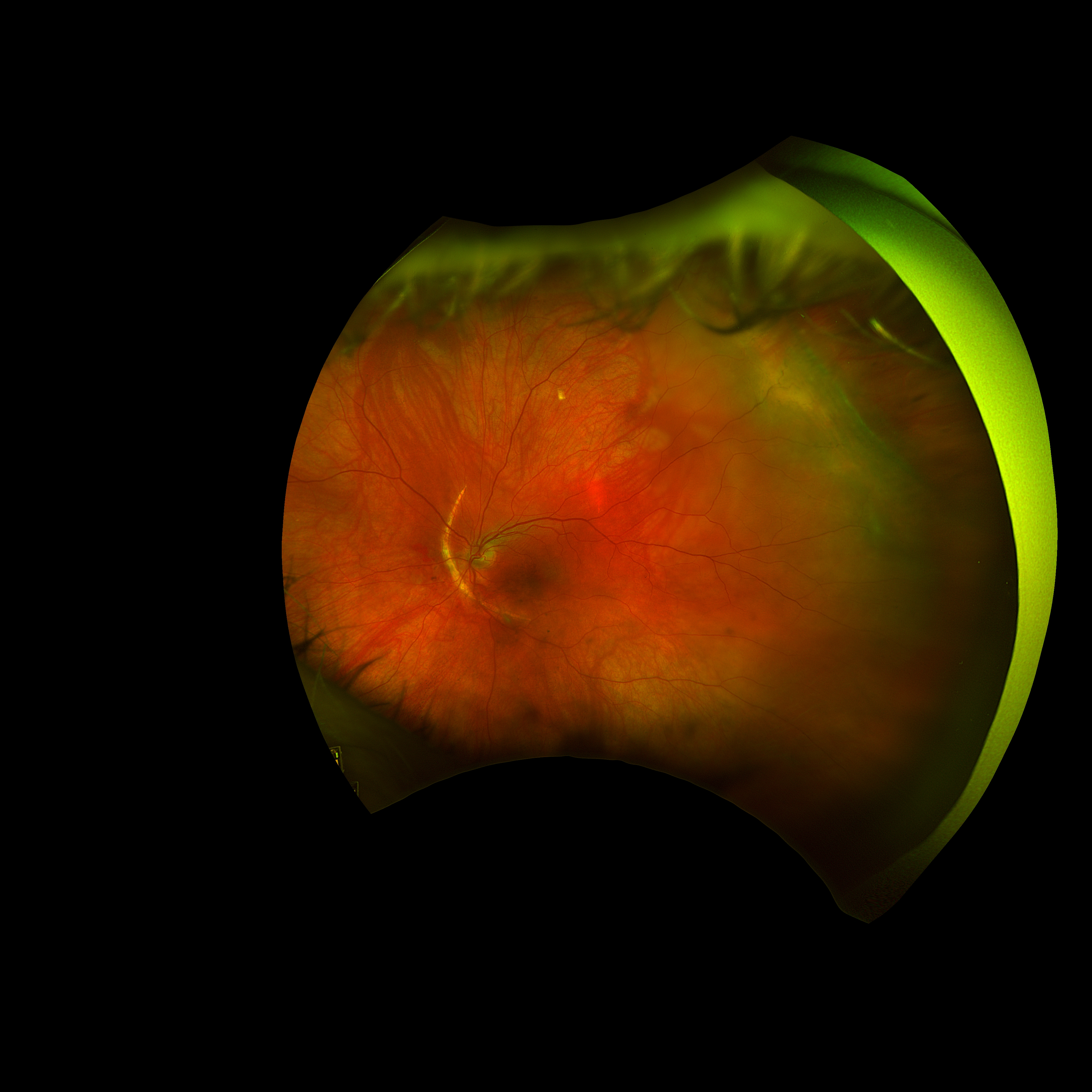
A 42-year-old woman sustained blunt trauma directly to her left globe after being kicked by a goat. On presentation, she was found to have multiple ocular injuries including a hyphema, ocular hypertension, iridodialysis, traumatic iritis, and vitreous hemorrhage. Upon clearance of her hyphema and vitreous hemorrhage after 14 days, she was found to have a retinal detachment and was referred to our center. Fundus examination demonstrated an arcuate choroidal rupture nasal to the optic nerve. There was a retinal dialysis from 2-4 o’clock with subretinal fluid from 1-5 o’clock that spared the macula. She underwent successful urgent retinal detachment repair with primary scleral buckle procedure.

Ophthalmic Atlas Images by EyeRounds.org, The University of Iowa are licensed under a Creative Commons Attribution-NonCommercial-NoDerivs 3.0 Unported License.


Address
University of IowaLegal
Related Links