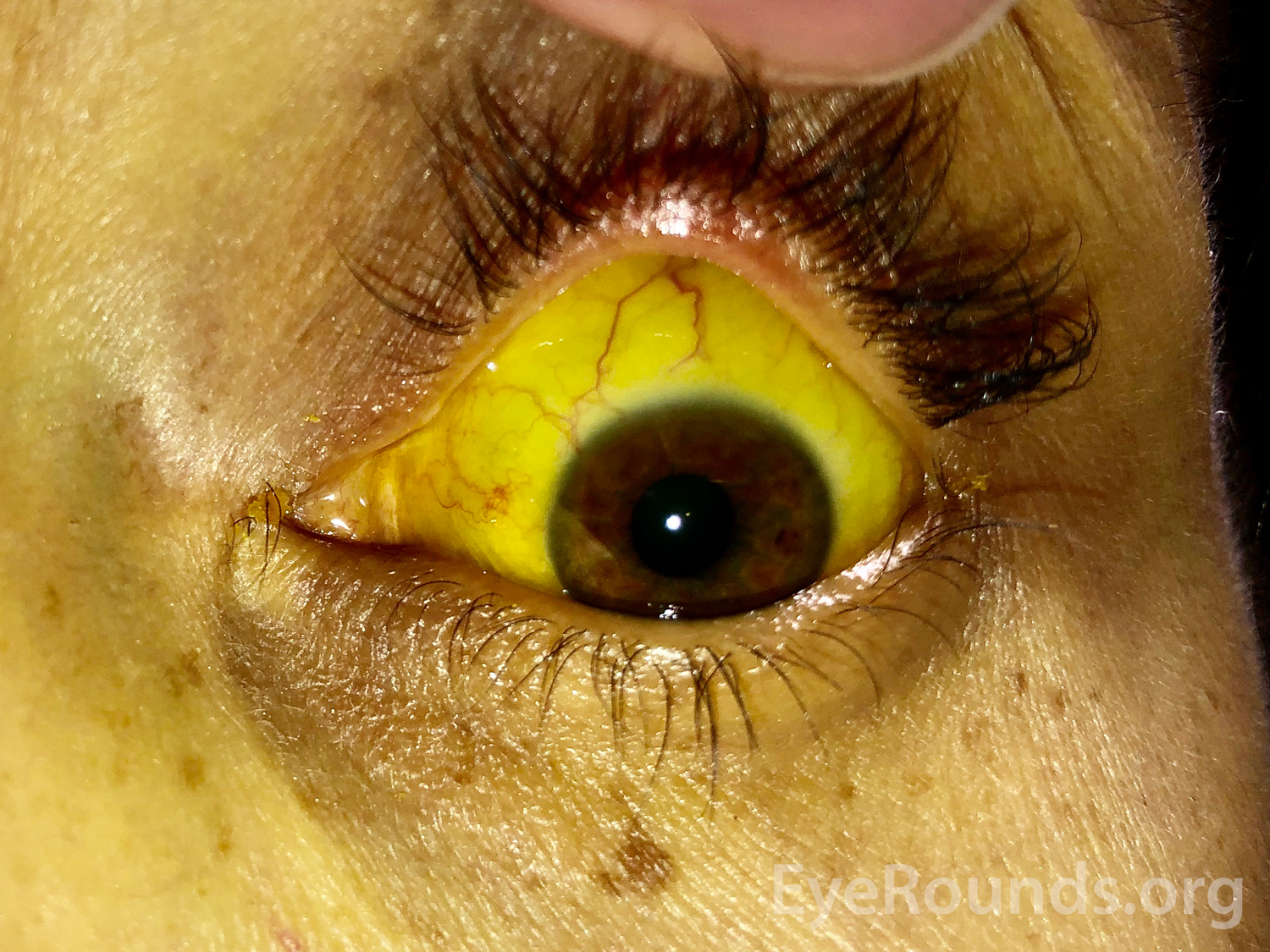
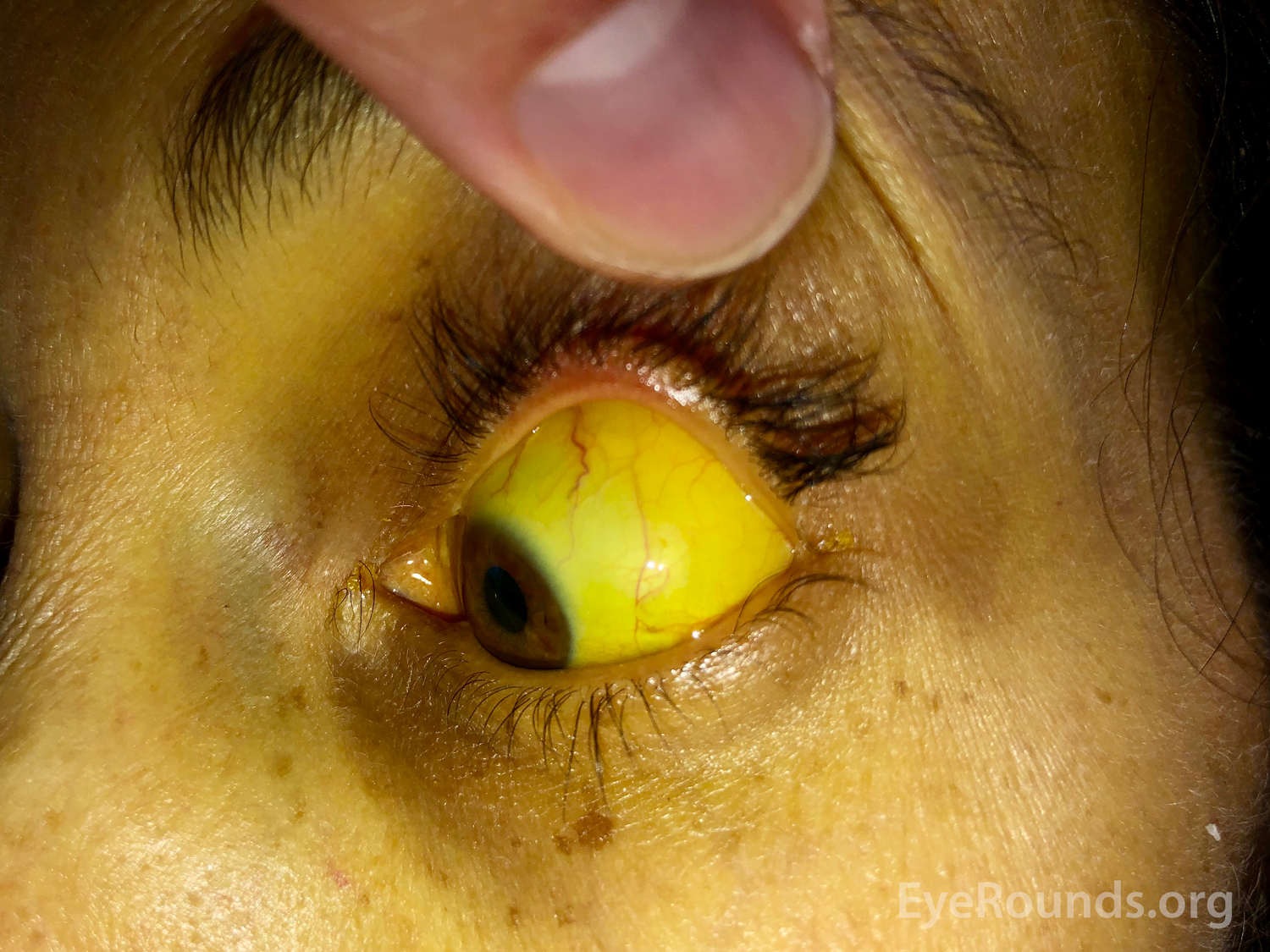
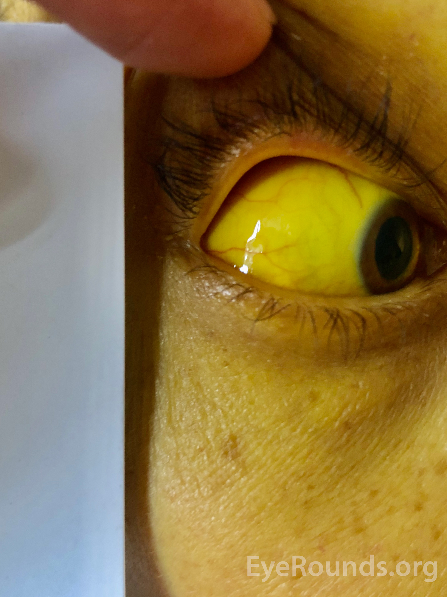
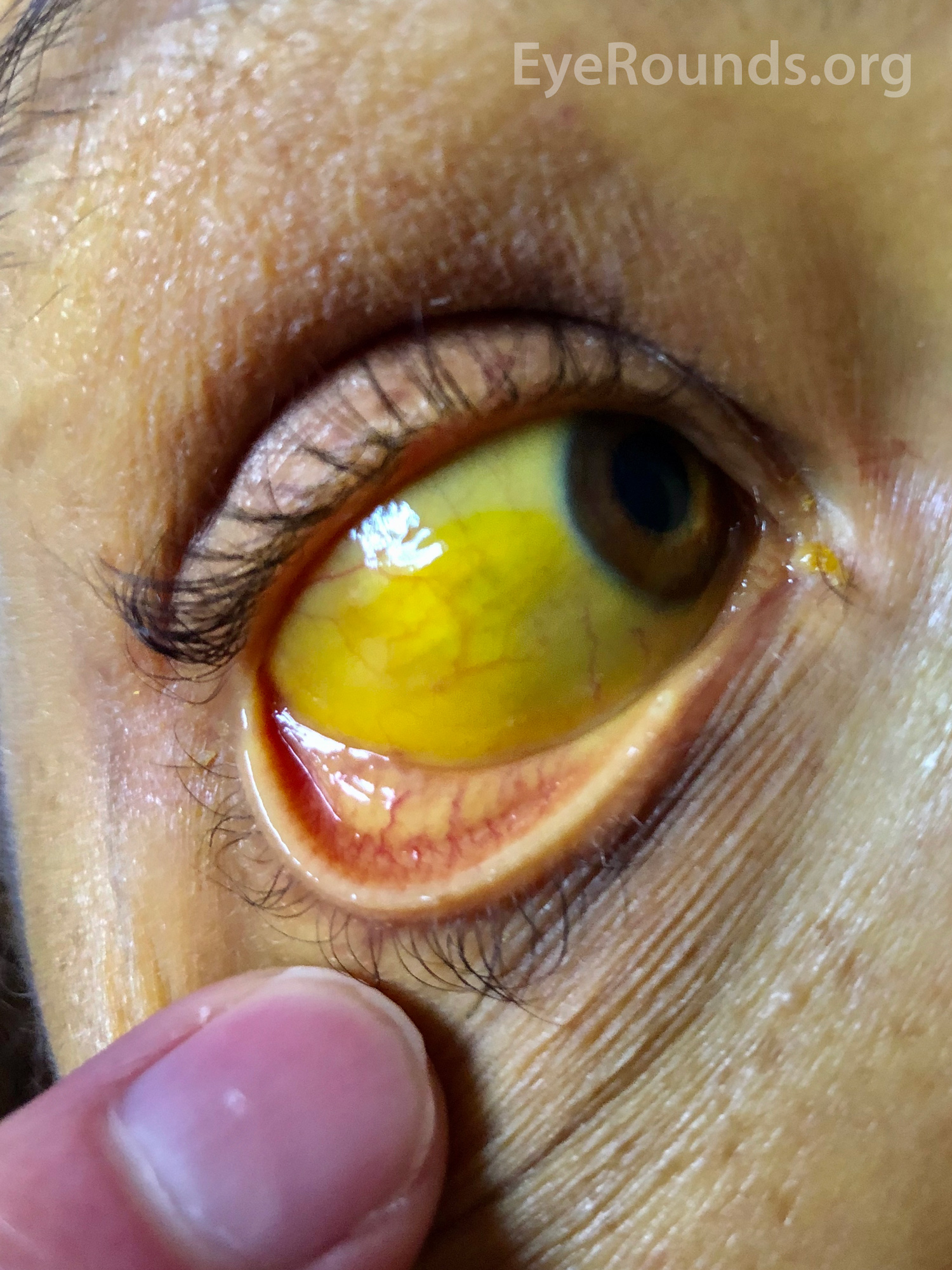
The image shows conjunctival icterus in a 49-year-old Hispanic female with decompensated alcoholic cirrhosis with severe alcoholic hepatitis. Bilirubin level is at 30 mg/dL (normal limit <=1.2 mg/dL). In addition to the yellowed conjunctiva, note the surrounding jaundiced skin. The yellow hue is better appreciated when seen in natural light and when juxtaposed with the white paper and/or the examiner's finger. The deposits of bilirubin are contained in the conjunctiva, and the color is accentuated by the white scleral background. Thus "scleral icterus" is technically an incorrect term.

Ophthalmic Atlas Images by EyeRounds.org, The University of Iowa are licensed under a Creative Commons Attribution-NonCommercial-NoDerivs 3.0 Unported License.


Address
University of IowaLegal
Related Links