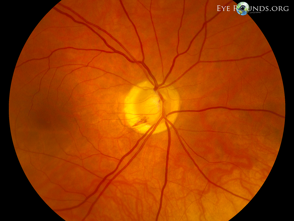
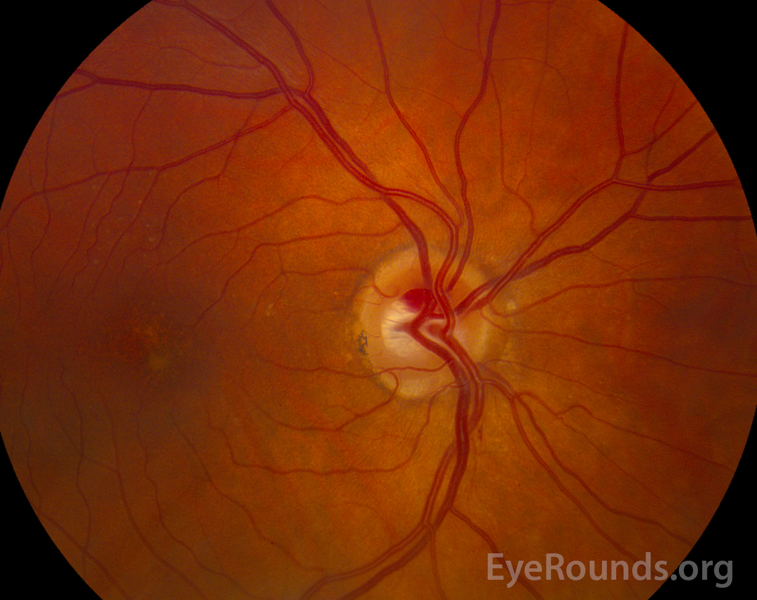
A 62-year-old female with primary open angle glaucoma was seen in the glaucoma clinic for follow-up and was incidentally found to have an intrapapillary hemorrhage in her right eye as shown below (figure 1). Intrapapillary hemorrhages are more common near the nasal border of the nerve in myopic eyes with tilted discs, and may be spontaneous or precipitated by acute disc edema, valsalva maneuver, or vitreopapillary traction [1]. The patient in this scenario was not myopic, and underwent echography to rule our optic nerve drusen or a vascular tumor. It was later revealed that she had been coughing recently, possible leading to a valsalva maneuver. These hemorrhages generally have a good prognosis and resolve spontaneously [1,2]. Of note, this hemorrhage is different than the typical peripapillary hemorrhage seen in normal tension glaucoma, which is located on the border of the border of the optic disc (see Optic Disc Hemorrhage in Normal Tension Glaucoma).

Ophthalmic Atlas Images by EyeRounds.org, The University of Iowa are licensed under a Creative Commons Attribution-NonCommercial-NoDerivs 3.0 Unported License.

Address
University of IowaLegal
Related Links