A 57-year-old female presented with chronic diabetic macular edema despite monthly and recent anti-VEGF therapy. The patient received 2 mg intravitreal triamcinolone because she was pseudophakic, had healthy optic nerves with normal intraocular pressure, and had no history of glaucoma.
Her vision in the affected right eye was 20/60, and her intraocular pressure was 14 by tonopen.
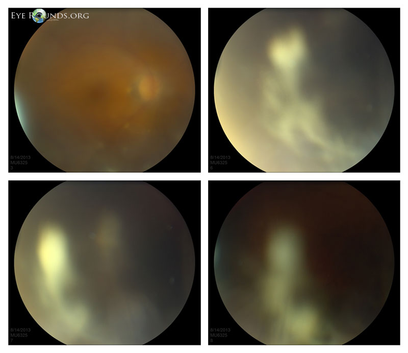
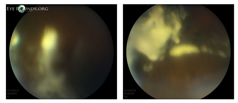
Dilated fundus exam of the right eye immediately following intravitreal injection of 2 mg (0.05 mL) triamcinolone in the inferotemporal quadrant. The view is hazy from the recent injection, as well as intentional focus in the vitreous. The yellow clumps represent triamcinolone in the vitreous cavity.
The purpose of these images is to demonstrate what triamcinolone looks like immediately following injection into the vitreous.
Triamcinolone acetonide (Kenalog) is a steroid that may be used for macular edema secondary to diabetes, vein occlusions, inflammatory conditions, etc.
The dose use varies, however 2 mg (or 0.05 mL) and 4 mg ( or 0.10 mL) are commonly used, studied, and well tolerated
Intravitreal triamcinolone is injected via a similar technique as anti-VEGF therapy; however, it is sometimes preferably injected inferotemporally due to its visibly apparent (non-transparent or “milky”) consistency. Also, a slightly larger gauge needle is sometimes needed (eg 27 or 30 gauge) due to larger particle size.
Preservative-free intravitreal Triesence (triamcinolone acetonide, Alcon) may also be used in a very similar fashion.
University of Iowa
Roy J. and Lucille A. Carver College of Medicine
Department of Ophthalmology and Visual Sciences
200 Hawkins Drive
Iowa City, IA 52242
University of Iowa
Roy J. and Lucille A. Carver College of Medicine
Department of Ophthalmology and Visual Sciences
200 Hawkins Drive
Iowa City, IA 52242