"Typical" iris colobomas are located in the inferonasal quadrant. They are caused by failure of the embryonic fissure to close in the 5th week of gestation, resulting in a "keyhole-shaped" pupil. They may be associated with colobomas of the ciliary body, choroid, retina, or optic nerve. "Atypical" iris colobomas are not caused by embryonic fissure closure defects and thus are not associated with other colobomas.
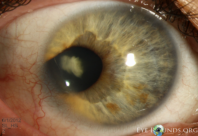
This patient presented with a temporal anterior segment coloboma in his right eye. This is unusual as colobomas are typically inferonasal, where the optic fissure fails to close completely during development.
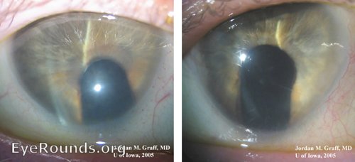
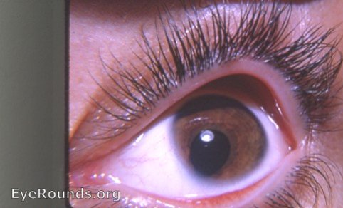
Typical coloboma of the iris, seen here as an inferonasal keyhole defect in the iris in both eyes (right and left eyes shown as if patient is facing the examiner with the nose removed).
Typical colobomas of the iris, as shown here, are caused by failure of the embryonic optic vessicle to close. The ciliary body, choroid, retina, and optic nerve may thus also be involved in patients with iris coloboma.
Iris colobomas can be associated with almost many types of chromosomal abnormalities and syndromes
*Dr. Caccamise has very generously shared his images of patients taken while operating during the "eye season" in rural India as well as those from his private practice during the 1960's and 1970's. Many of his images are significant for their historical perspective and for techniques and conditions seen in settings in undeveloped areas.
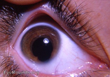
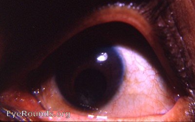
Coloboma of iris: the bridge variety
The pyriform shape of the microcornea together with its pointing in the direction of the fetal fissure indicates that a coloboma of the iris is present. The coloboma is in a special category, i.e. the bridge coloboma. Through computer manipulation, an attempt has been made to make visible the thin bridge of iris tissue that crosses the coloboma gap to complete the pupil in that area. One must look very carefully to see this bridge. Of course, with the slit-lamp it is readily visible.
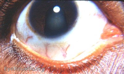
The patient appeared with what appeared to be a large " complete " sector iridectomy inferiorly. Slight central and posterior subcapsular cataractous changes were evident - congenital. The patient had no memory of surgery. The shape and size of the coloboma and the normal shape of the cornea suggested that this was a surgical coloboma - not a developmental, congenital coloboma.
Coloboma of the iris is frequently bilateral. The pyriform shape of the cornea is a usual accompaniment of the uveal defect. The pear-shape points to the fetal fissure, i.e. inferonasally. The 3rd photo suggests a surgical iridectomy - in spite of a denial of surgery - because these features of a congenital coloboma are missing.
This photograph is classic for congenital coloboma of the iris. Whenever the cornea presents with this pear-shape and is pointed inferonasally, there is a strong possibility that there will be a keyhole coloboma of the iris in that area.
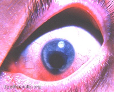
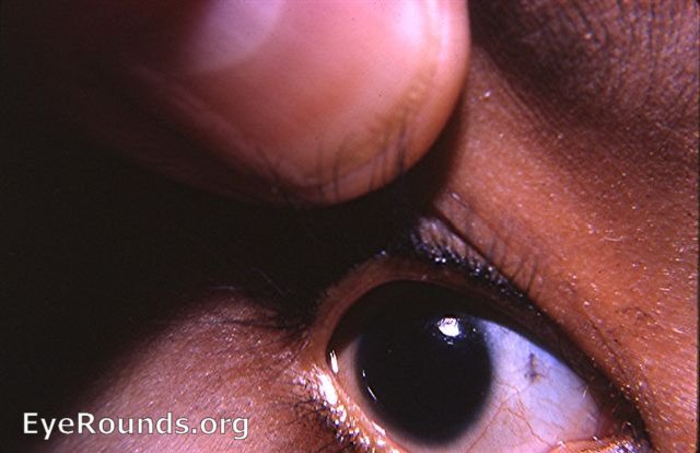
Congenital coloboma of the iris with pyriform cornea
The pyriform nature with pointing inferiorly should immediately suggest a congenital coloboma of the iris inferiorly at the location of the fetal fissure. The coloboma in this eye cannot be seen in this photograph because the iris is so dark brown. Fundus examination at times may reveal varying extension of the uveal defect posteriorly - even to the point of including the optic nerve. The vertical pigmented line a few mms from the cornea is due to pigmentation at the points of normal bloodvessel perforation of the sclera in this heavily pigmented patient. Patients with coloboma of the iris should be examined carefully for other developmental defects.
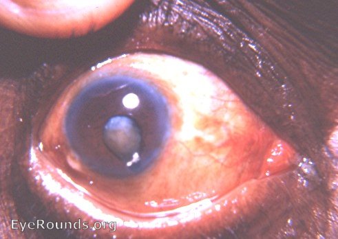
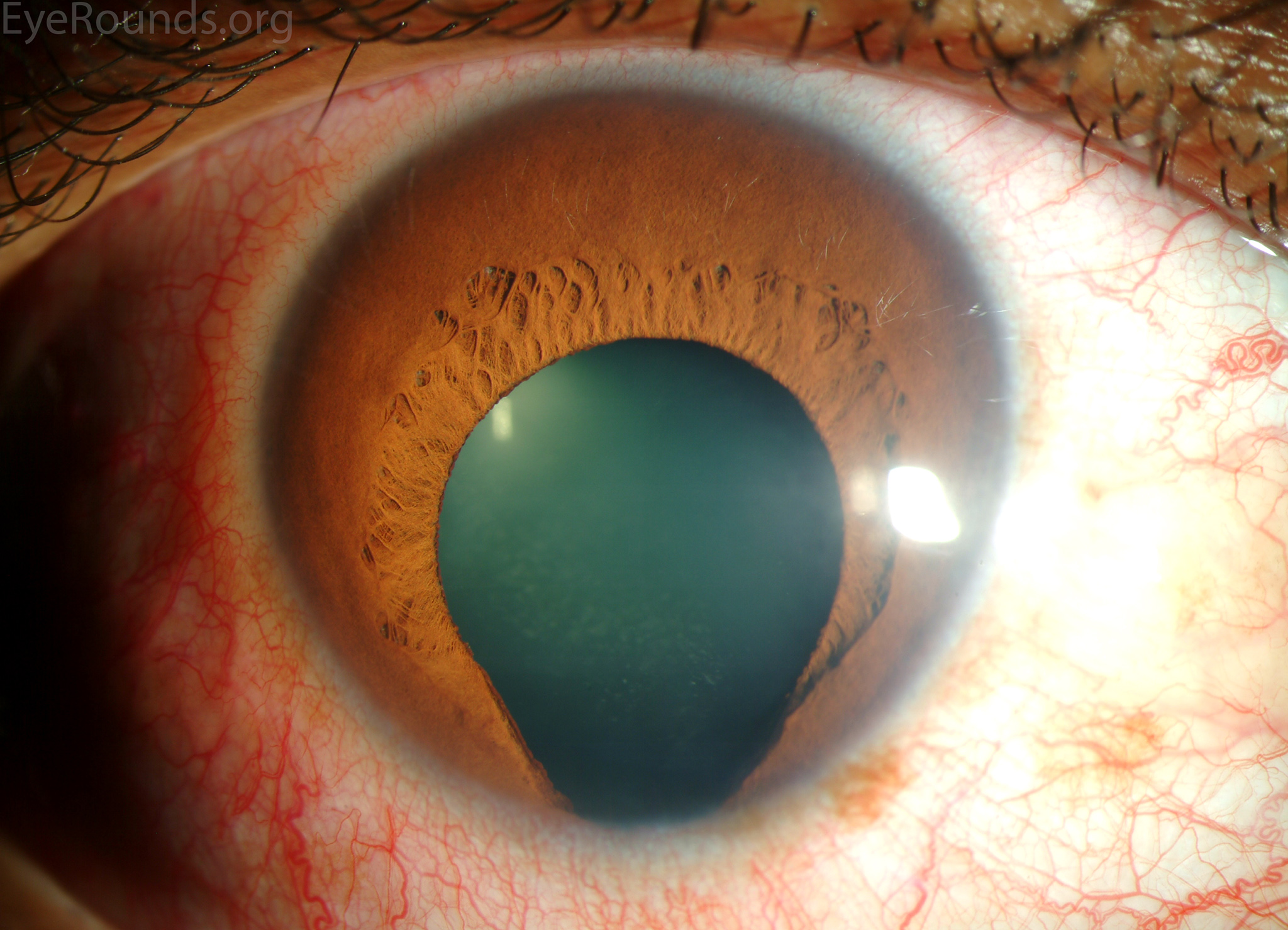
Coloboma of iris - a special bridge variety-with a mature corticonuclear cataract
A spedial type of congenital coloboma of the iris is the bridge coloboma.From Duane-Fuchs: " In this the pupil is separated from the coloboma by a narrow thread of iris tissue, which stretches like a bridge from one pillar of the coloboma to the other." The cataract is mature because the entire lens is cataractous - something that is rarely attained by a cataract undergoing surgery in America now. The layman's question about his cataract and surgery used to be : "is my cataract ripe " Ripe meant mature - ready for surgery. In India the word for ripe was "pukka".
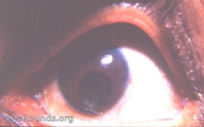
Computer manipulation of Congenital coloboma of iris with immature cortical cataract
Computer manipulation decreases quality but allows the inferior coloboma to be seen vaguely. A cataract is seen in the inferior portion of the coloboma.
University of Iowa
Roy J. and Lucille A. Carver College of Medicine
Department of Ophthalmology and Visual Sciences
200 Hawkins Drive
Iowa City, IA 52242
University of Iowa
Roy J. and Lucille A. Carver College of Medicine
Department of Ophthalmology and Visual Sciences
200 Hawkins Drive
Iowa City, IA 52242