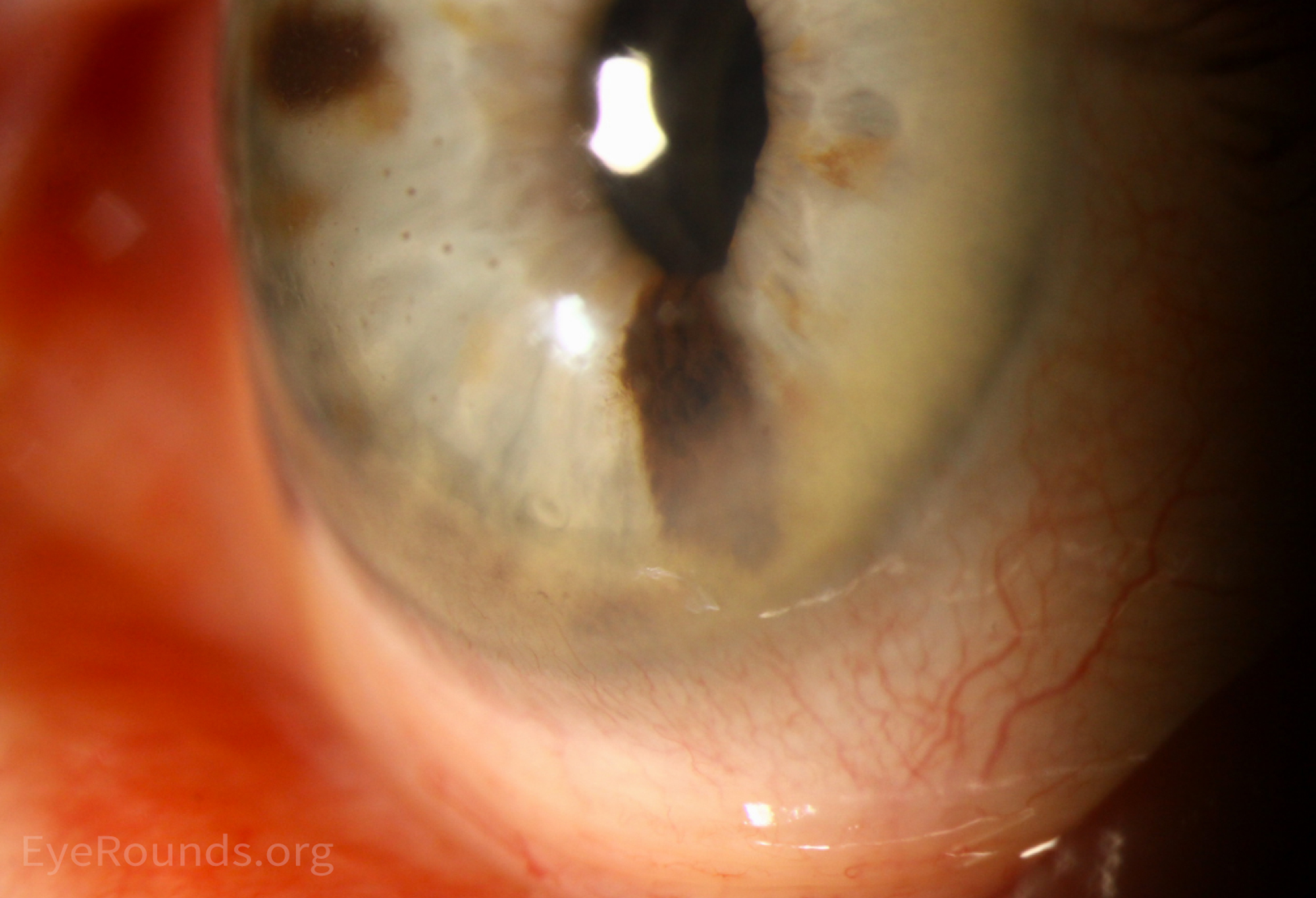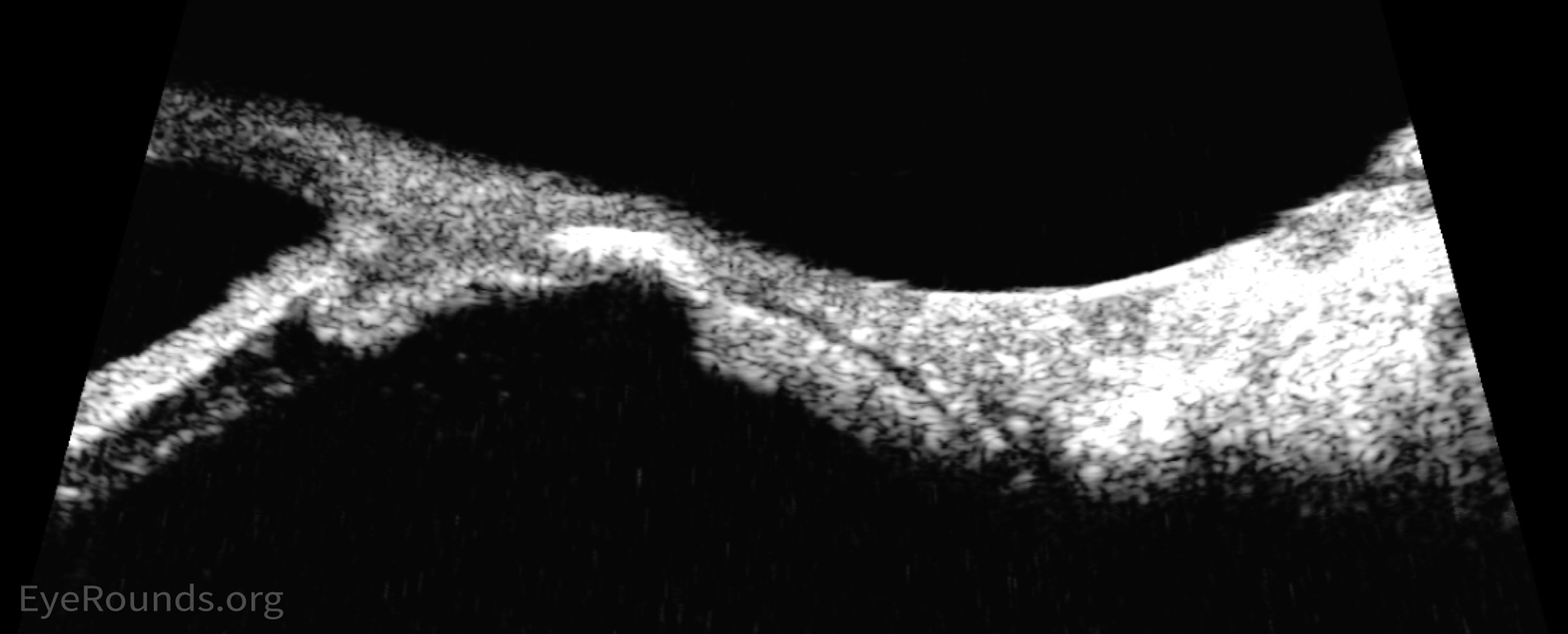
93-year-old woman presented with progression of longstanding iris nevus. Her original presentation in 2005 is shown in Figure 1. Figures 2 and 3 demonstrate frank enlargement of the primary lesion at 6 o'clock and local metastasis, with erosion through the angle into the sclera at 8o'clock (Figures 4 and 5). Pigmented keratic precipitates are suspicious for anterior chamber seeding (Figure 6). High frequency anterior segment imaging demonstrates further angle invasion (arc of high reflectivity; Figure 7).


3 patients with iris melanomas. The last patient had a recurrence 20 years later after an iridocyclectomy.
81-year-old female with a slowly enlarging iris lesion found to be an iris melanoma originating from an iris nevus.

Ophthalmic Atlas Images by EyeRounds.org, The University of Iowa are licensed under a Creative Commons Attribution-NonCommercial-NoDerivs 3.0 Unported License.