EyeRounds Online Atlas of Ophthalmology
Contributor: William Charles Caccamise, Sr, MD, Retired Clinical Assistant Professor of Ophthalmology, University of Rochester School of Medicine and Dentistry
*Dr. Caccamise has very generously shared his images of patients taken while operating during the "eye season" in rural India as well as those from his private practice during the 1960's and 1970's. Many of his images are significant for their historical perspective and for techniques and conditions seen in settings in undeveloped areas.
Membranous Cataracts
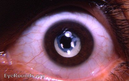
Congenital cataract in the membranous cataract stage

This photo was taken by Dr. Caccamise in 1967. The patient was an adult Caucasian who had no history of eye surgery. Under full mydriasis, this membranous cataract was found. It demonstrates the degree of self-absorption that a cataract can undergo without surgery. Refer to the discussion that accompanies the other membranous cataract photo in this Atlas.
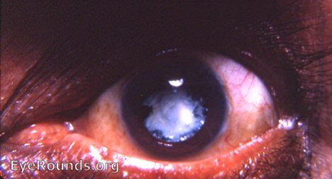
Cataracta membranacea

Although this initially appeared to be a dense aftercataract (the German Nachstar), there was no history of cataract surgery and there was no evidence of a von Graefe knife incision superiorly. There was no evidence of an iridectomy. There was no evidence of a point of entrance of a needling knife. Therefore, the conclusion was that this represented a membranous cataract.This type of cataract is a rarity even in rural India. It is unlikely to be experienced by opthalmologists practicing solely in America. When it does occur, it is usually in a child who had a congenital cataract or one that appeared in early childhood when no hard nucleus was involved. The development of this membranous cataract involves inspissation, disintegration, and resorption. In the photo, calcification has occurred in some areas. Black spots in the white membrane represent relatively transparent areas in the membranous cataract.
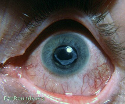
Remnant of congenital cataract through self-absorption

This membrane is the remnant of a congenital cataract that self-absorbed. No cataract surgery of any type was ever performed on this eye.

Ophthalmic Atlas Images by EyeRounds.org, The University of Iowa are licensed under a Creative Commons Attribution-NonCommercial-NoDerivs 3.0 Unported License.


