Microcornea is defined as a cornea less than 10 mm in diameter. It is thought that microcornea occurs secondary to an arrest in corneal development due to overgrowth of the tips of the optic cup. The differential diagnosis includes cornea plana, sclerocornea, nanophthalmos, and anterior microphthalmos.
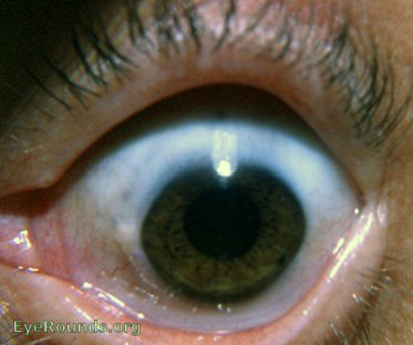
The patient in this photograph has microcornea (corneal diameter 5 mm) associated with primary congenital glaucoma. The condition is associated with an increased risk of glaucoma and patients are usually hyperopic.
Contributor: William Charles Caccamise, Sr, MD, Retired Clinical Assistant Professor of Ophthalmology, University of Rochester School of Medicine and Dentistry
*Dr. Caccamise has very generously shared his images of patients taken while operating during the "eye season" in rural India as well as those from his private practice during the 1960's and 1970's. Many of his images are significant for their historical perspective and for techniques and conditions seen in settings in undeveloped areas.
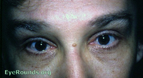
Microcornea, both eyes
Except for the microcornea, no other general or ocular abnormality was evident. In microcornea, the measurement is less than 11 mm in the greatest diameter.In this patient, the right cornea was slightly smaller than the left cornea - both were smaller than normal.
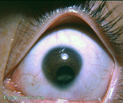
The cornea measured 9.5mm in diameter. A measurement less than 11mm is considered microcornea. In this case, it is part of a microphthalmos. The word nanophthalmos is a synonym for microphthalmos.
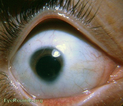
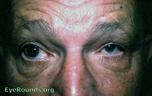
Microcornea is a cornea that measures <11mm. This cornea measures 10mm in diameter. The eye is normal except for slight microphthalmos. As an added observation: pingueculae are located nasally and temporally.
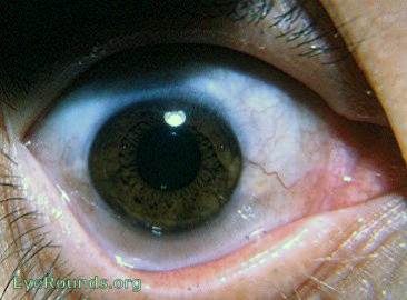
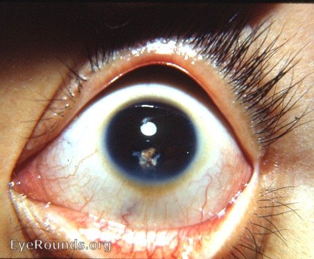
The microcornea presents the pear-shape that always suggests the possibility of a congenital coloboma of the iris. Cataractous changes are evident in the lens inferiorly.
The left cornea meets the standard for microcornea, i.e. a diameter < 11mm. The right cornea is borderline for megalocornea, i.e.>13mm. There is heterochromia: the right iris is brown, the left iris is bluish.
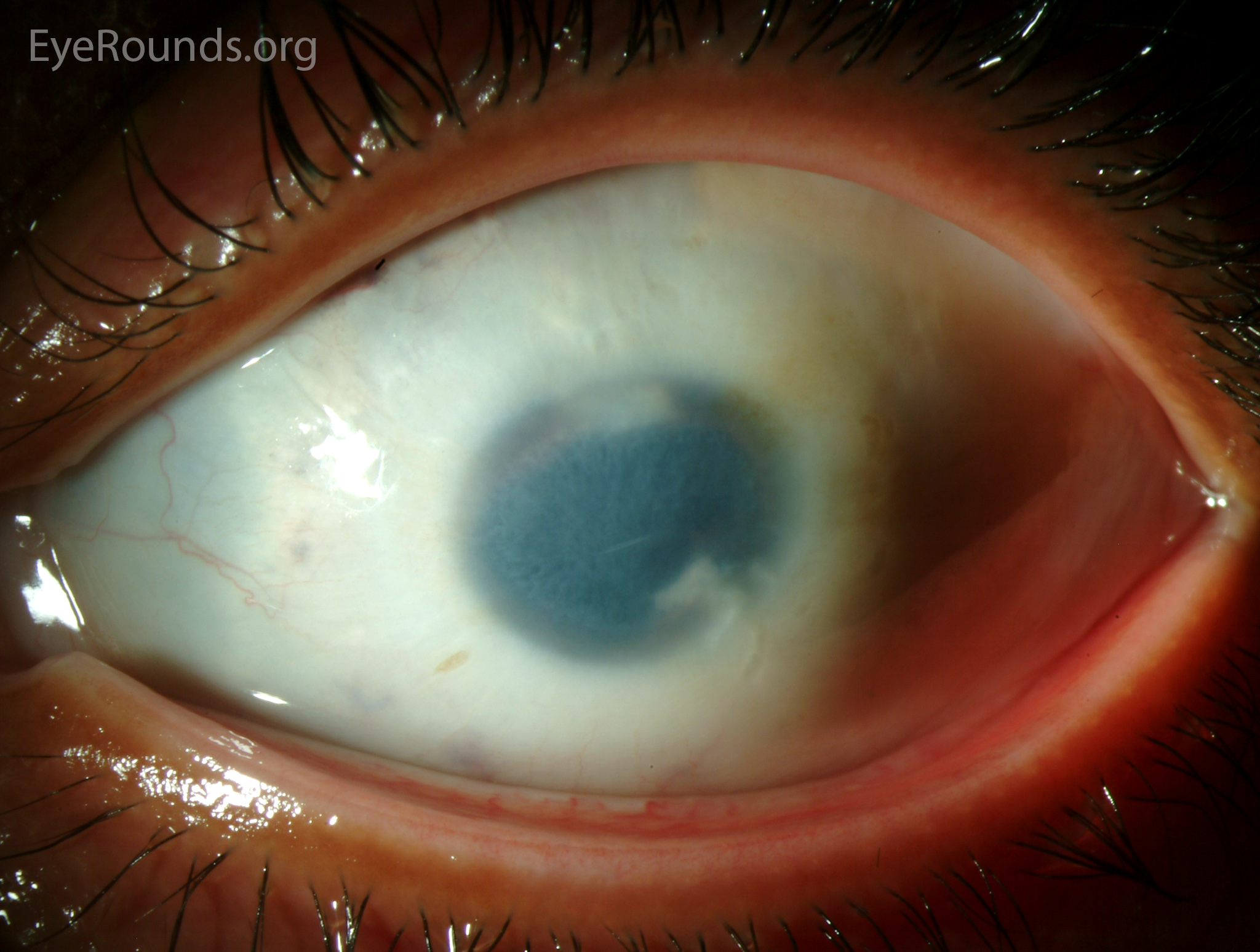
Microcornea with after-cataract following needling of congenital cataract
This 10-year-old Tibetan girl was seen in consultaion at the Eye Clinic. The history was that of a congenital cataract in the left eye. Needling had been done. A significant after-cataract effectively obstructed the pupil. During the evaluation a diagnosis of congenital microcornea was made.A corneal diameter less than 11.0 mm is termed microcornea. The cornea was normal except for its small size.However, microcornea may be associated with other abnormalities, viz. congenital cataract as in this eye. Incidentally, megalocornea indicates a corneal diameter greater than 13 mm.
Narumi Y, Nishina S, Tokimitsu M, et al. Identification of a novel missense mutation of MAF in a Japanese family with congenital cataract by whole exome sequencing: a clinical report and review of literature. Am J Med Genet A. 2014;164A(5):1272-6.
Nischal KK. Corneal abnormalities. Pediatric ophthalmology and strabismus 2e. New York: Springer; 2002;391-429.
University of Iowa
Roy J. and Lucille A. Carver College of Medicine
Department of Ophthalmology and Visual Sciences
200 Hawkins Drive
Iowa City, IA 52242
University of Iowa
Roy J. and Lucille A. Carver College of Medicine
Department of Ophthalmology and Visual Sciences
200 Hawkins Drive
Iowa City, IA 52242