

EyeRounds Online Atlas of Ophthalmology
Contributor: William Charles Caccamise, Sr, MD, Retired Clinical Assistant Professor of Ophthalmology, University of Rochester School of Medicine and Dentistry
*Dr. Caccamise has very generously shared his images of patients taken while operating during the "eye season" in rural India as well as those from his private practice during the 1960's and 1970's. Many of his images are significant for their historical perspective and for techniques and conditions seen in settings in undeveloped areas.
Category: External Disease
Nodular episcleritis
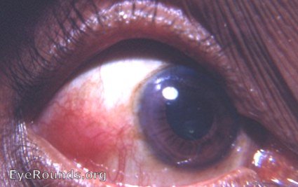
The differential diagnosis is between nodular episcleritis and nodular scleritis. Nodular episcleritis occurs as a localized, painful, inflammatory, episcleral nodule as seen in the photo. Note: Nodular scleritis is more extensive and deeper.
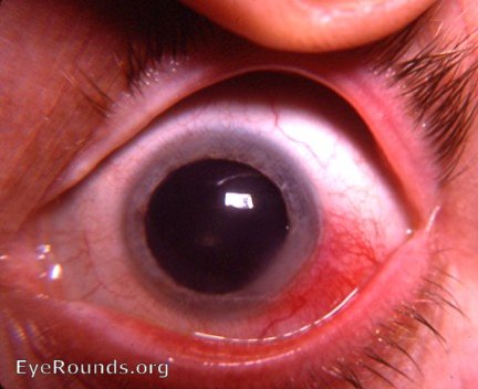
Here a diagnosis of nodular episcleritis was made - on healing there should be no residual of scleral damage
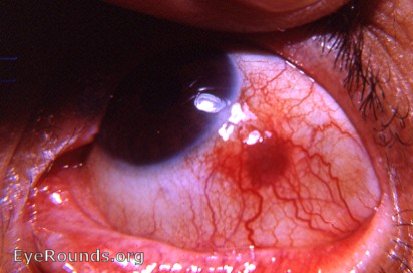 the initial more active stage of nodular episcleritis - nodular scleritis
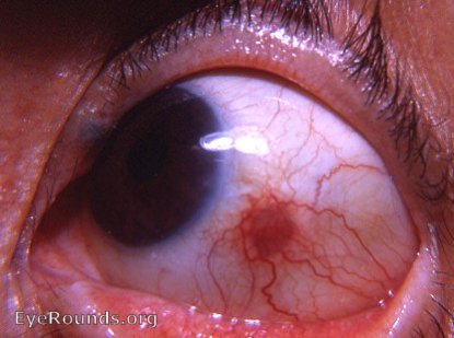
This photo shows the healing phase of what appears to be nodular episcleritis. If scleral damage is evident when the lesion has finally healed, the diagnosis of nodular scleritis will have to be entertained.

Ophthalmic Atlas Images by EyeRounds.org, The University of Iowa are licensed under a Creative Commons Attribution-NonCommercial-NoDerivs 3.0 Unported License.