This site uses tracking information. Visit our privacy policy. Click to agree to this policy and not see this again.

Ophthalmology and Visual Sciences

Oil droplet cataract

Oil droplet cataract

Category(ies): Cataract, Lens
Contributor: Andrew Doan, MD, PhD

The patient was a 57-year-old man with vision loss in the right eye (OD) greater than left eye (OS) in the setting of a craniopharyngioma, which was resected 16 years prior. His visual acuity had been no light perception OD and 20/20 OS since the procedure.

Retinoscopy shows oil droplet cataract
45-year-old with slow, progressive loss of vision in both eyes (20/200). Pinholes to 20/25 but cannot be refracted. Retinoscopy shows oil droplet cataract. Slit lamp shows a black, empty space between lens cortex and lens nucleus. Difficult to see without looking at the red reflex.

Oil droplet cataract

Oil droplet cataract

Category(ies): Cataract, Lens
Contributors: Peter H. Sanchez, MD, and Ryan J. Diel, MD
Photographer: Alex Steggall

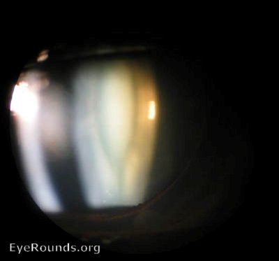
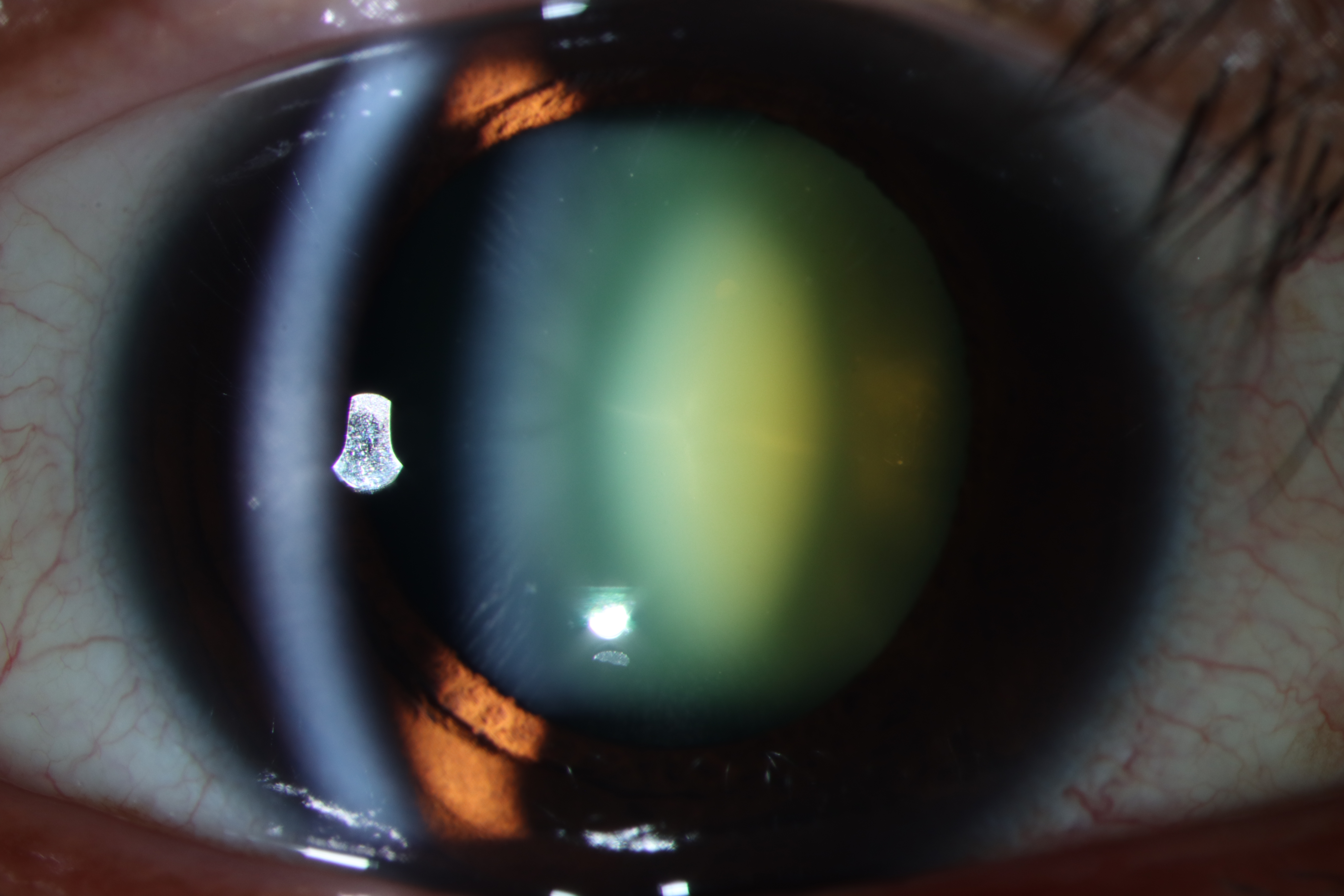
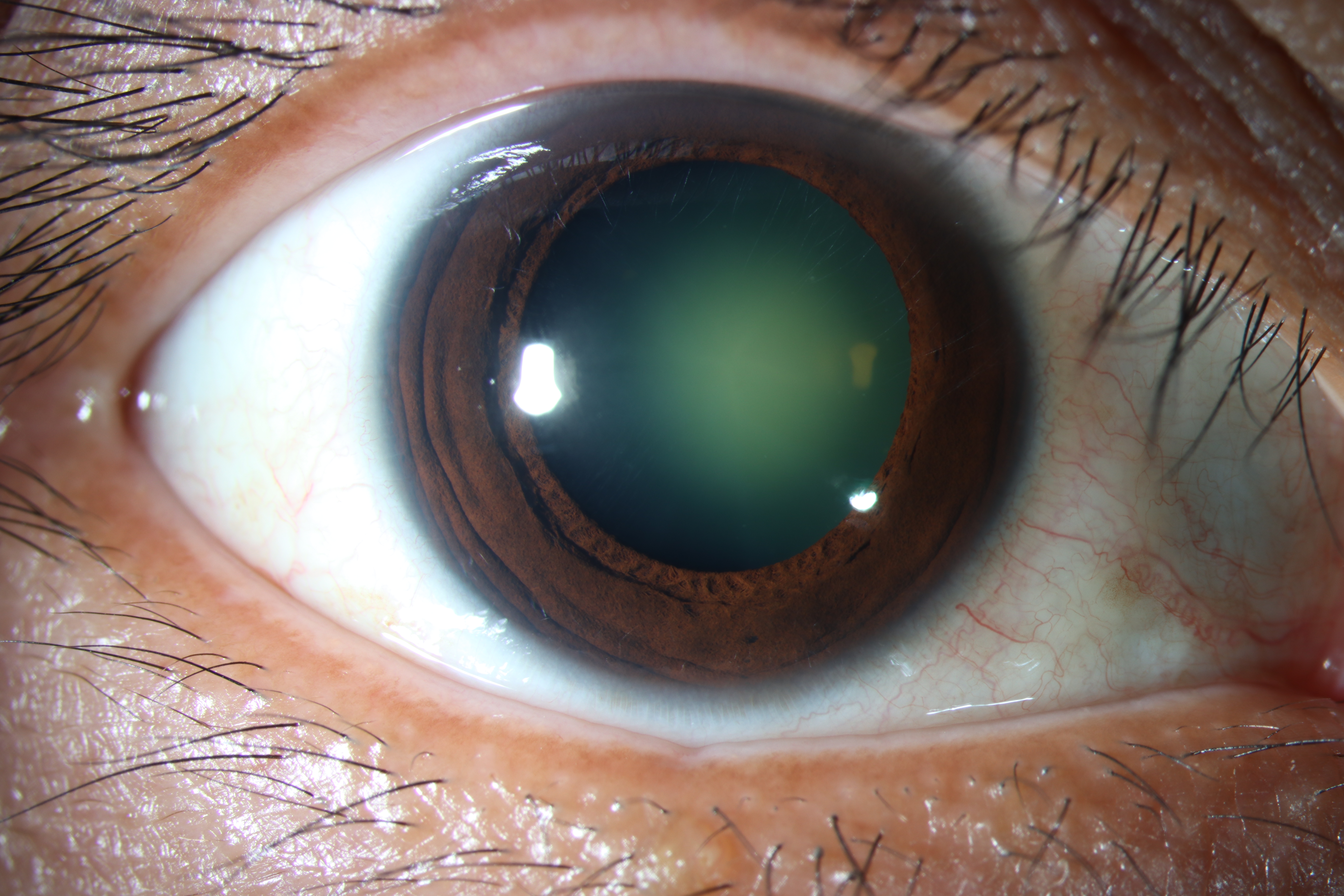
39 y/o female presented for evaluation of blurry vision in the right eye and difficulties with glare at night. She denied any history of ocular trauma, diabetes, or steroid use. Her uncorrected visual acuity OD was 20/1250 with improvement to 20/200 with pinhole. Uncorrected OS was 20/20-1. Her subjective refraction was -15.50 +1.00 x 137 OD (20/150) and -0.50 +0.50 x 090 OS (20/20). Her exam was notable for a 3+ oil-droplet cataract OD. Lens-calculations were performed and showed axial eye-lengths of 23.05 mm OD and 22.94 mm OS. She was offered cataract extraction with intraocular lens implantation.

Diffuse illumination slit lamp photograph of an oil-droplet cataract showing central nuclear yellow opacity of the lens with dark clearing surrounding the oil-droplet.
Optical section slit lamp photograph of the right eye showing a 3+ oil-droplet cataract with the central yellow opacified nucleus surrounded by an area of dark clearing followed by the cortical and capsular layers anteriorly.