This site uses tracking information. Visit our privacy policy. Click to agree to this policy and not see this again.

Ophthalmology and Visual Sciences

Choroidal osteoma

Choroidal osteoma

Category(ies): Retina, Vitreous
Contributor: Jesse Vislisel, MD
Photographer: Brice Critser, CRA

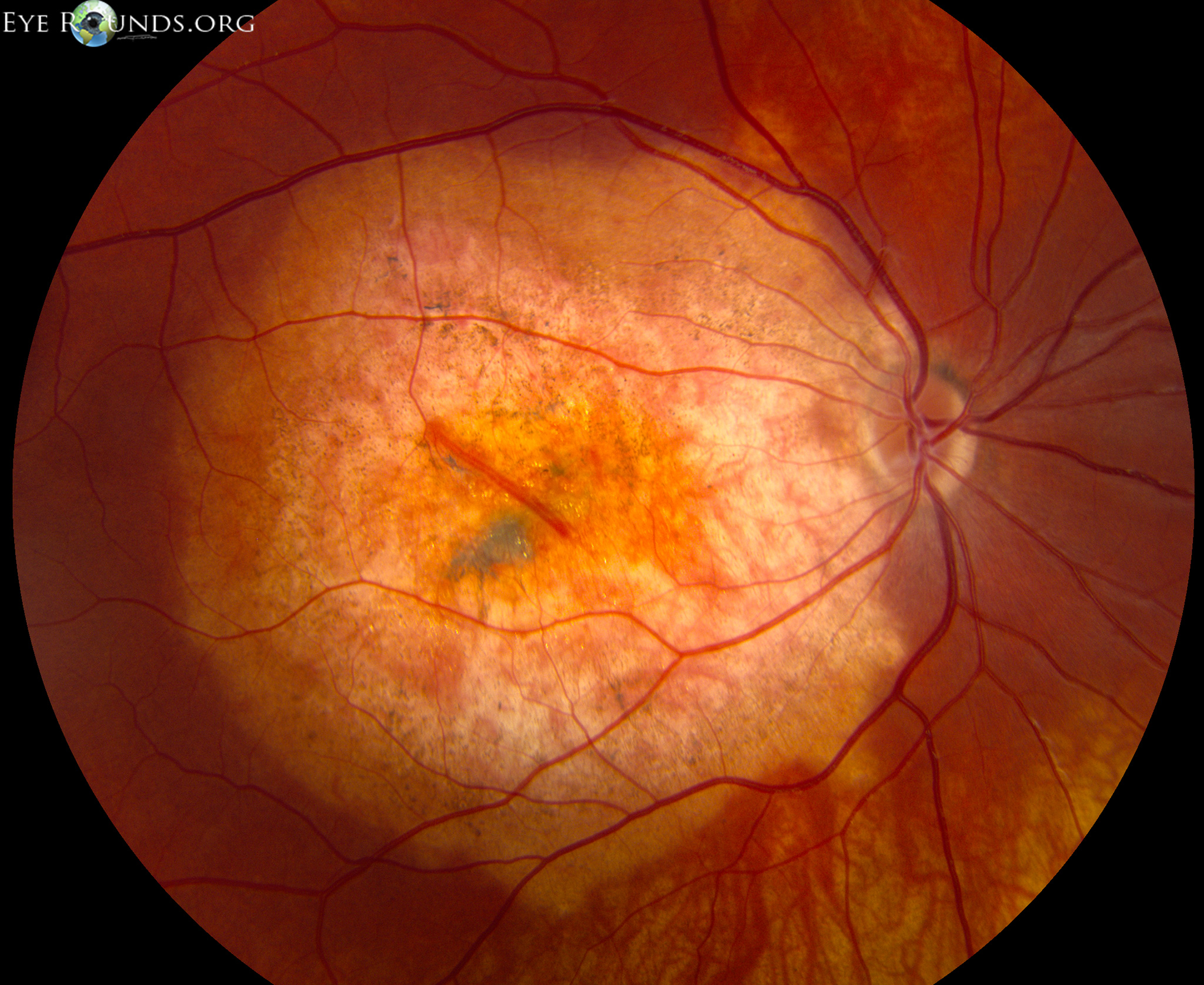
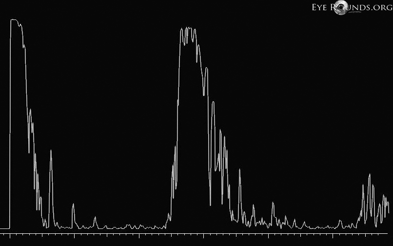
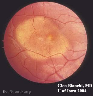
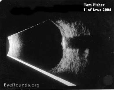
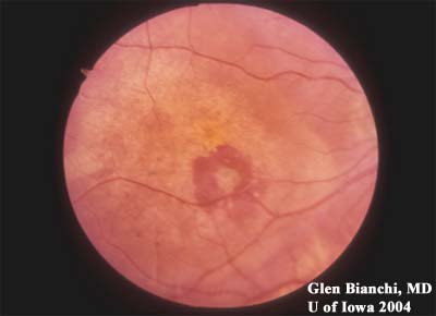
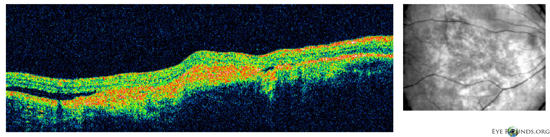
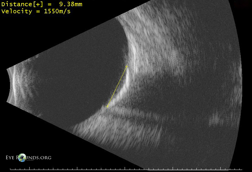
Choroidal osteomas are benign neoplasms in which areas of the choroid are replaced with mature bone. The tumors are most commonly solitary, unilateral, and juxtapapillary in location which helps differentiate them from idiopathic sclerochoroidal calcification. They appear clinically as ovoid subretinal lesions, ranging from yellow-white to orange-red in color, sometimes with overlying pigmentary changes.

fundus image choroidal osteoma
An individual with a choroidal osteoma beneath the macula in the right eye.
OCT image choroidal osteoma
OCT scan portrays a highly-reflective lesion at the level of the choroid with associated subretinal fluid.
A-scan image choroidal osteoma
The A-scan shows the high reflectivity of the mass.
A-scan image choroidal osteoma
The B-scan reveals the extent of the highly echogenic lesion and the associated acoustic shadowing which is characteristic for these tumors.

Other choroidal osteoma images

Other choroidal osteoma images

Contributor: Glen Bianchi, MD and Andrew Doan, MD, PhD, University of Iowa

Other choroidal osteoma images

Other choroidal osteoma images

Contributor: Andrew Doan, MD, PhD, and Tom Fisher, University of Iowa
Alternative fundus choroidal osteoma
Note shadowing on B-scan