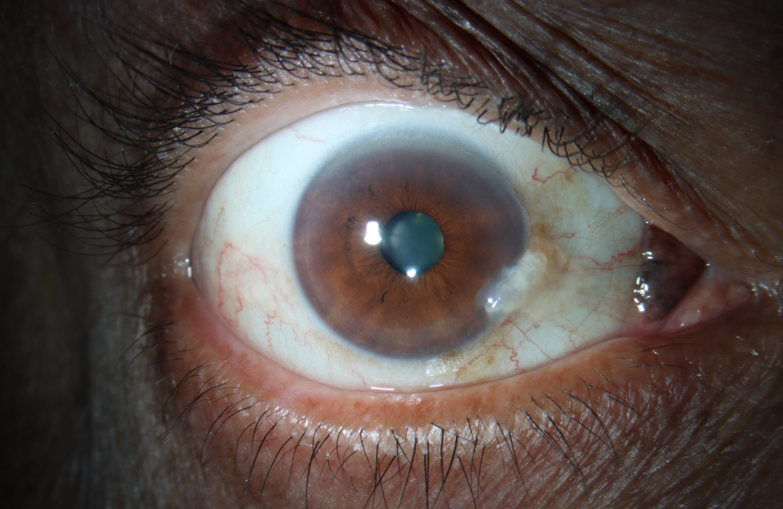
A 63-year-old man presented for a conjunctival lesion he says has been present his whole life but has been concerning to his daughter in law. Although his history was reassuring, its location near the caruncle as well as his immunosuppressed status warranted excisional biopsy. Histologically, sections demonstrated nests and sheets of nevus cells variably demonstrating cytoplasmic pigment. A diagnosis of a plica semilunaris nevus was made.

Ophthalmic Atlas Images by EyeRounds.org, The University of Iowa are licensed under a Creative Commons Attribution-NonCommercial-NoDerivs 3.0 Unported License.


Address
University of IowaLegal
Related Links