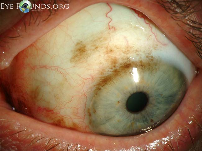
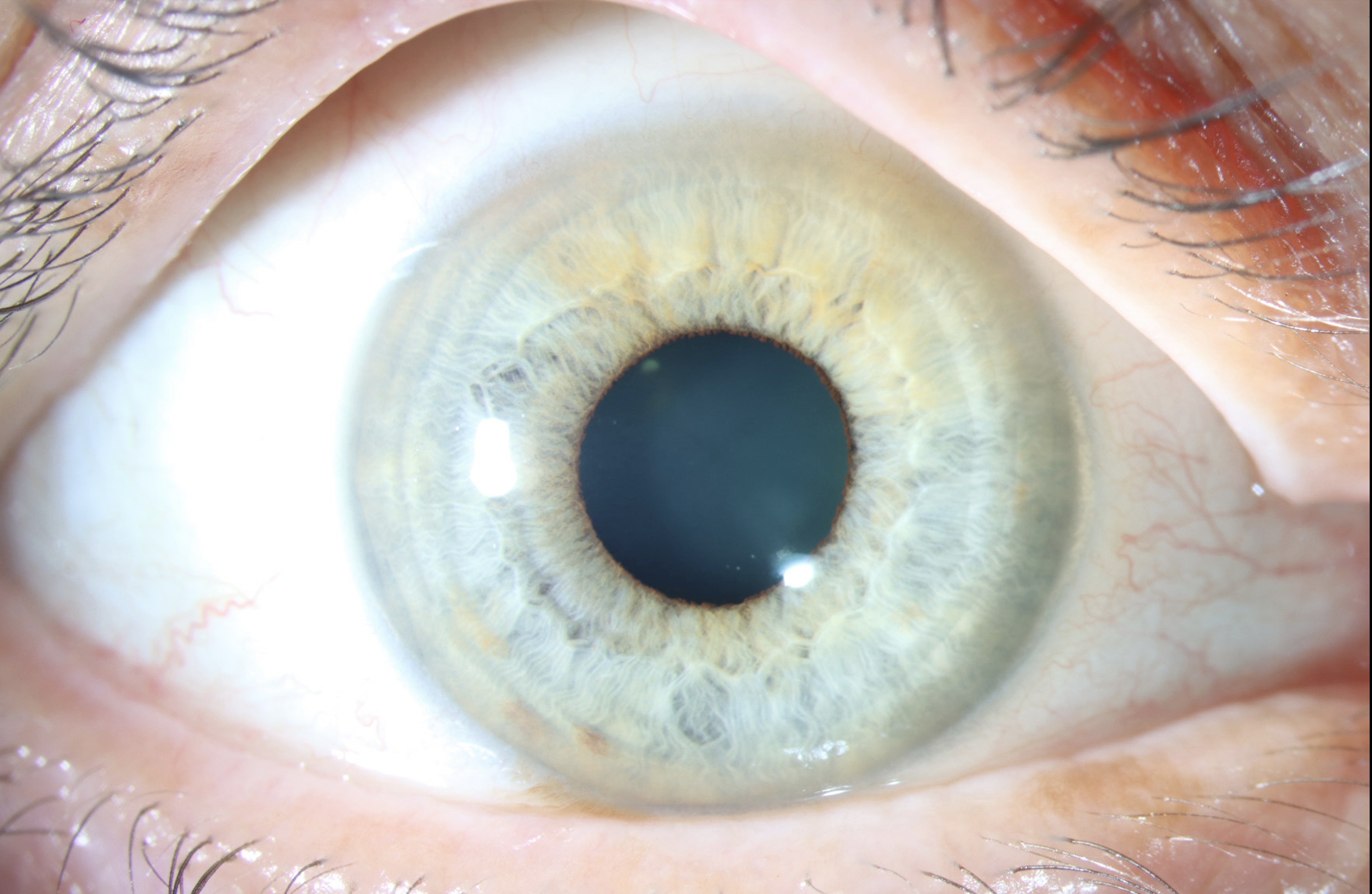
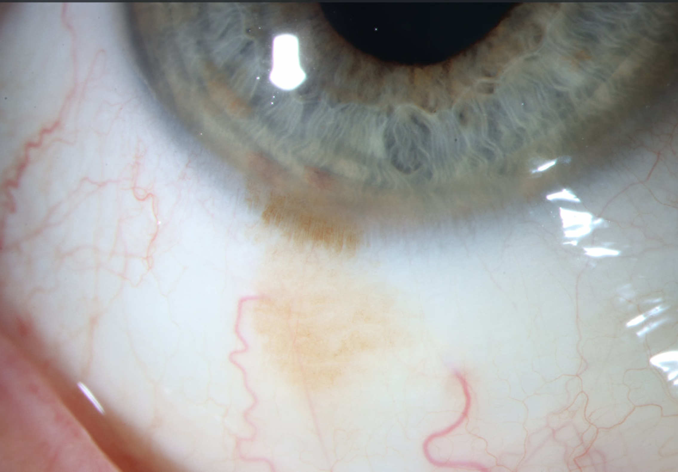
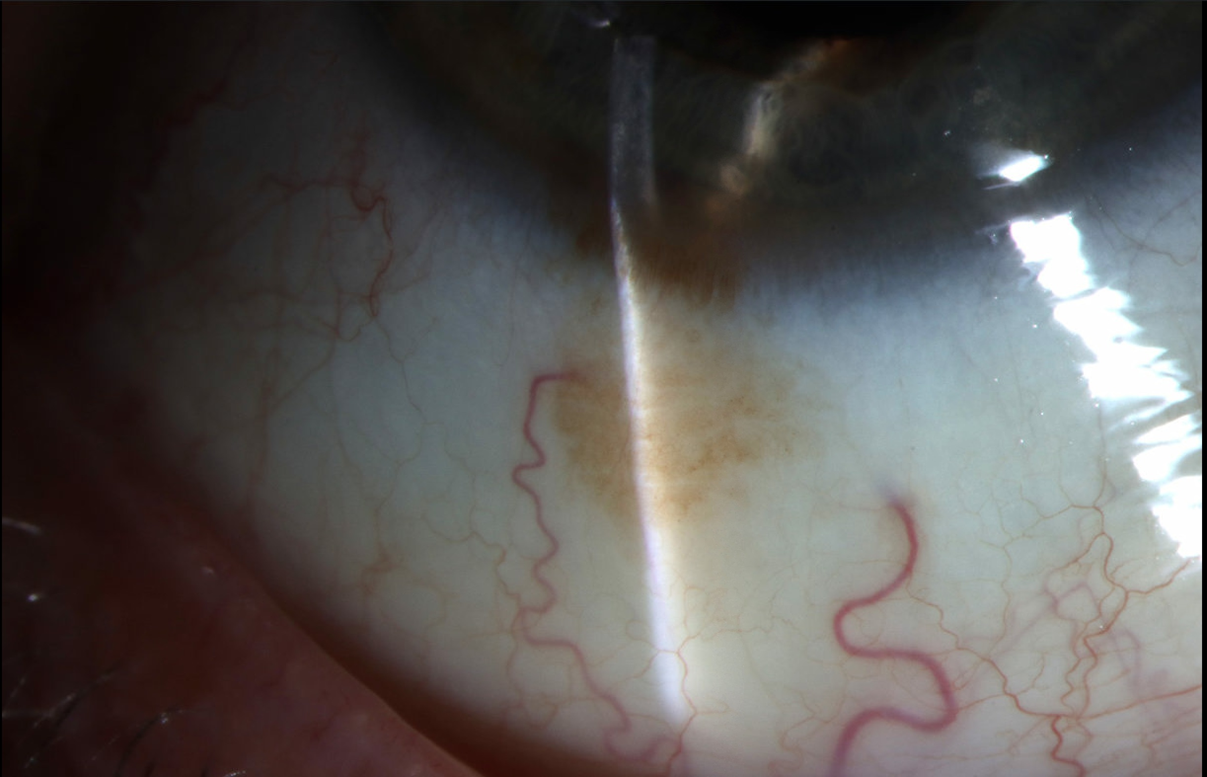
Primary acquired melanosis (PAM) is an acquired pigmentation of the conjunctival epithelium. It is analagous to lentigo maligna of the skin and carries a risk of malignant transformation. PAM appears as unilateral, patchy areas of flat, brown pigmentation within the superficial conjunctiva. The areas may wax and wane over time.
Slit lamp photos of the right eye from a 66-year-old Caucasian man with one clock hour of flat brown pigmented conjunctival tissue at the inferior bulbar quadrant that extends into the limbus. No feeder vessels were identified. Gonioscopy showed no evidence of ciliary body tumor. The patient first noticed this spot two weeks prior to presentation. These findings were consistent with primary acquired melanosis (PAM). Two small iris nevi were also seen inferiorly, above the area of PAM. A small lower lid margin nevus measuring 1.5 mm was identified nasally on the same eye. As no concerning features were identified on examination, annual surveillance including repeat slit lamp photography was advised to monitor for progression.

Ophthalmic Atlas Images by EyeRounds.org, The University of Iowa are licensed under a Creative Commons Attribution-NonCommercial-NoDerivs 3.0 Unported License.


Address
University of IowaLegal
Related Links