This site uses tracking information. Visit our privacy policy. Click to agree to this policy and not see this again.

Ophthalmology and Visual Sciences

Pyogenic granuloma

Pyogenic granuloma

Category(ies): Cornea, External Eye Disease
Contributor: David A. Ramirez, MD, Heather A. Stiff, MD, Mark A. Greiner, MD
Photographer:Jody Troyer, CRA

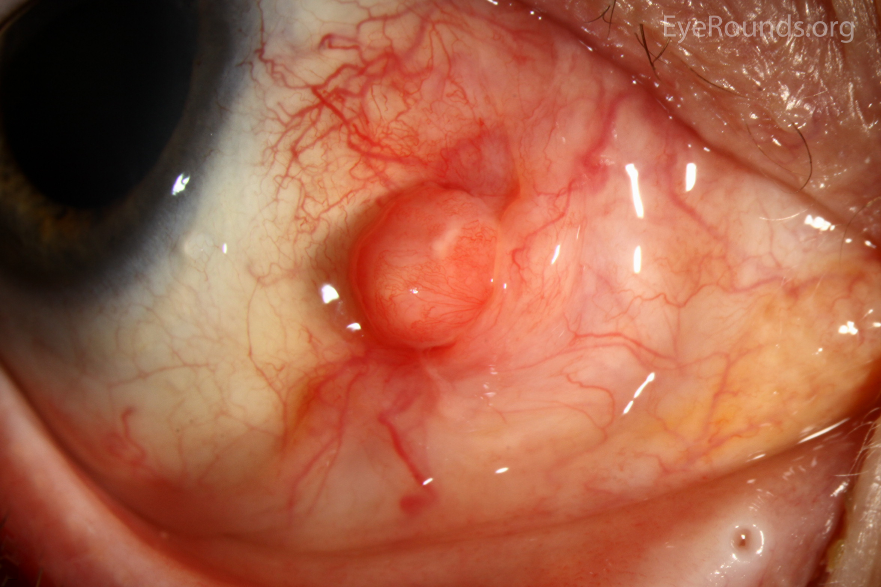
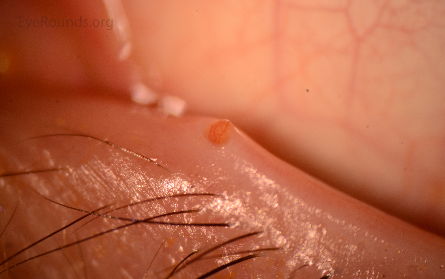
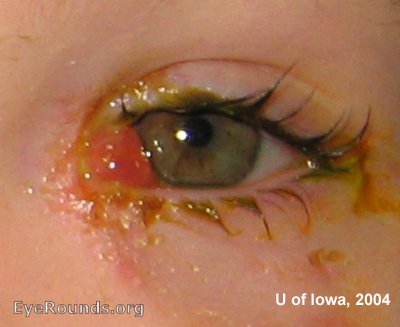
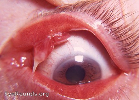
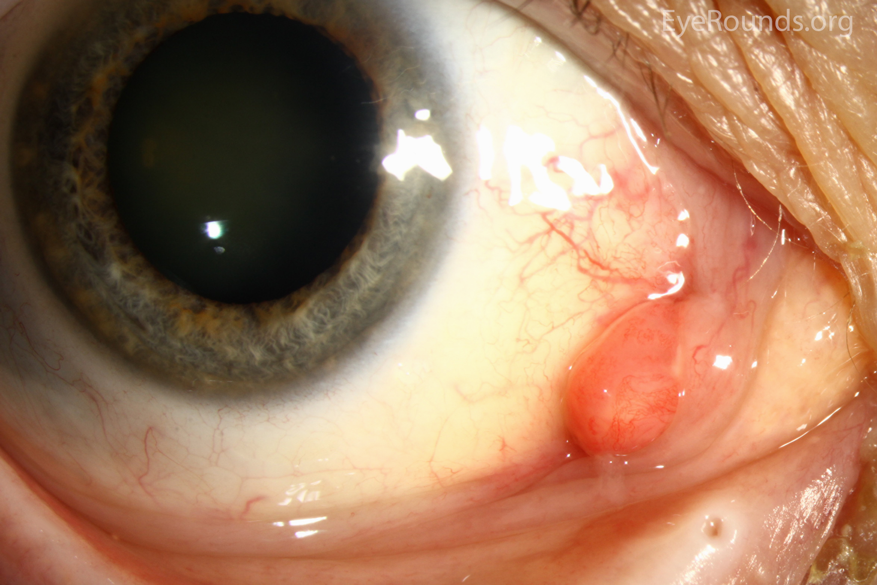
A pyogenic granuloma is characterized as a pedunculated vascular lesion, and appears as a fleshy red or pink mass. It is often located on the eyelid and bleeds easily with minimal contact. Clinically, it arises with rapid growth in an area of previous trauma, surgery, or inflammation. Histologically, it is composed of granulation tissue, capillaries, and non-granulomatous inflammatory cells [1 ,2]. Recommended treatment is excision of the lesion at it's base.

Pyogenic granulomas slit lamp
Figure 1. Slit lamp photograph of the left eye shows a nasal bulbar pyogenic granuloma with surrounding conjunctival injection. Pathology showed that underlying changes were consistent with pterygium or pingueculum. The overlying area of acute hemorrhage and fibrosis was thought to be due to digital trauma from rubbing.

Contributor: Jesse Vislisel, MD
Photographer: Antoinette Venckus, CRA

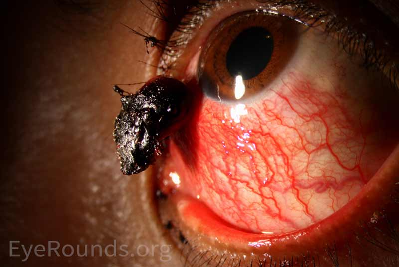
Pyogenic granulomas are formed when there is abnormally vigorous proliferation of granulation tissue. They appear as vascular growths with smooth surfaces, usually at the site of previous ocular surgery.

Pyogenic granulomas
Figure 2: The patient in these photographs developed this lesion after undergoing a scleral buckle procedure for the repair of a retinal detachment. The growth was later excised and pathology findings were consistent with pyogenic granuloma.
Pyogenic granulomas enlarged
Figure 2: The patient in these photographs developed this lesion after undergoing a scleral buckle procedure for the repair of a retinal detachment. The growth was later excised and pathology findings were consistent with pyogenic granuloma.

Contributor: Tressa Larson, OD, FAAO
Photographer: Mark Greiner, MD
Pyogenic granulomas
Figure 3: These photos are from a patient who had previously had intracanalicular punctal plugs.
Pyogenic granulomas enlarged
Figure 3: These photos are from a patient who had previously had intracanalicular punctal plugs.

Contributor: Tressa Larson, OD, FAAO
Photographer: Mark Greiner, MD
Pyogenic granulomas
Figure 4: This lesion occurred a few months after undergoing canalicular repair. The lesion was surgically excised and the pathology specimen showed extensive granulation tissue with parenchymal hemorrhage.
Reference:
  • Mannis MJ, Holland, Edward J. Cornea; Fundamentals, Diagnosis, and Management. Fourth ed: Elsevier, 2017.
  • Neff A, Chahal H, Carter KD. Benign Eyelid Lesions Ophthalmology. Fifth ed: Elsevier 2019.

Image Permissions:
Creative Commons

Ophthalmic Atlas Images by EyeRounds.org, The University of Iowa are licensed under a Creative Commons Attribution-NonCommercial-NoDerivs 3.0 Unported License.