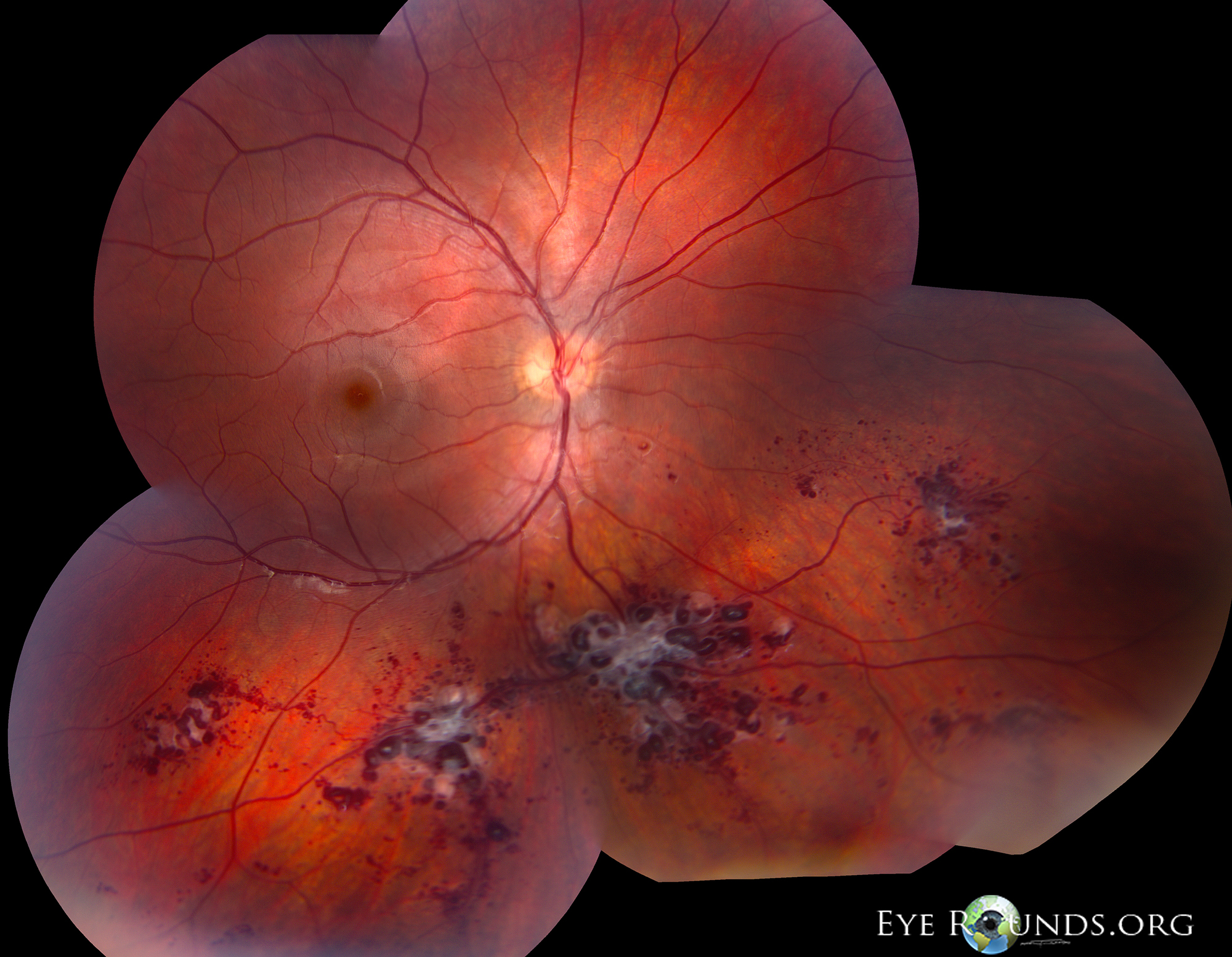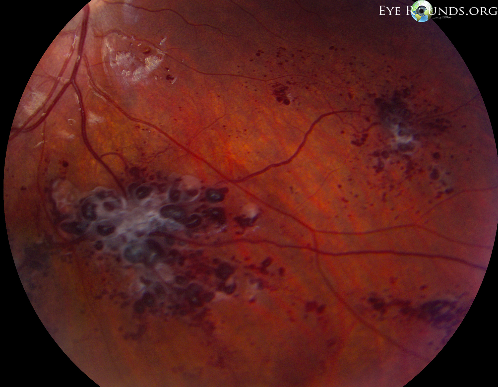
9-year-old boy with an unremarkable birth and childhood history, presented with a history of incidental findings suggestive of retinal cavernous hemangiomas in the right eye seen during routine optometric exam. He has no history of patching. No visual complaints, and no history of trauma.
Of note, his father has a history of many cavernous hemangiomas involving the brain, with associated headaches and seizures. His paternal grandmother also had intracranial and liver cavernous hemangiomas.
BCVA cc: OD 20/20; OS 20/20
IOP: 15 and 20
SLE: unremarkable, both eyes (OU)


The left eye, not shown, was unremarkable. The optic nerve had a 0.1 cup-to-disc ratio and a healthy pink rim. The macula was flat and dry, and the vessels were normal. The periphery was intact. There were no vitreous opacities or heme. No PVD or cells.
Upon further work-up with pediatric neurology and neurosurgery, this patient was found to have multiple small intracranial hemangiomas, including in the cerebellum.
Retinal cavernous hemangiomas are rare vascular hamartomas, composed of dark intraretinal aneurysms with characteristic "cluster-of-grapes" appearance.
Usually unilateral and rarely increase in size.
Almost always asymptomatic, although macular involvement or vitreous hemorrhage has been reported.
Skin and central nervous system hemangiomas may co-exist, and autosomal dominant pedigrees have been reported. Genotyping for three known cerebral cavernous malformation, or CCM, genes can establish a hereditary cause.
Caution: do not confuse this with retinal capillary hemangiomas (i.e. discrete tumors with feeder vessels) which is classically seen with von Hippel Lindau disease.

Ophthalmic Atlas Images by EyeRounds.org, The University of Iowa are licensed under a Creative Commons Attribution-NonCommercial-NoDerivs 3.0 Unported License.