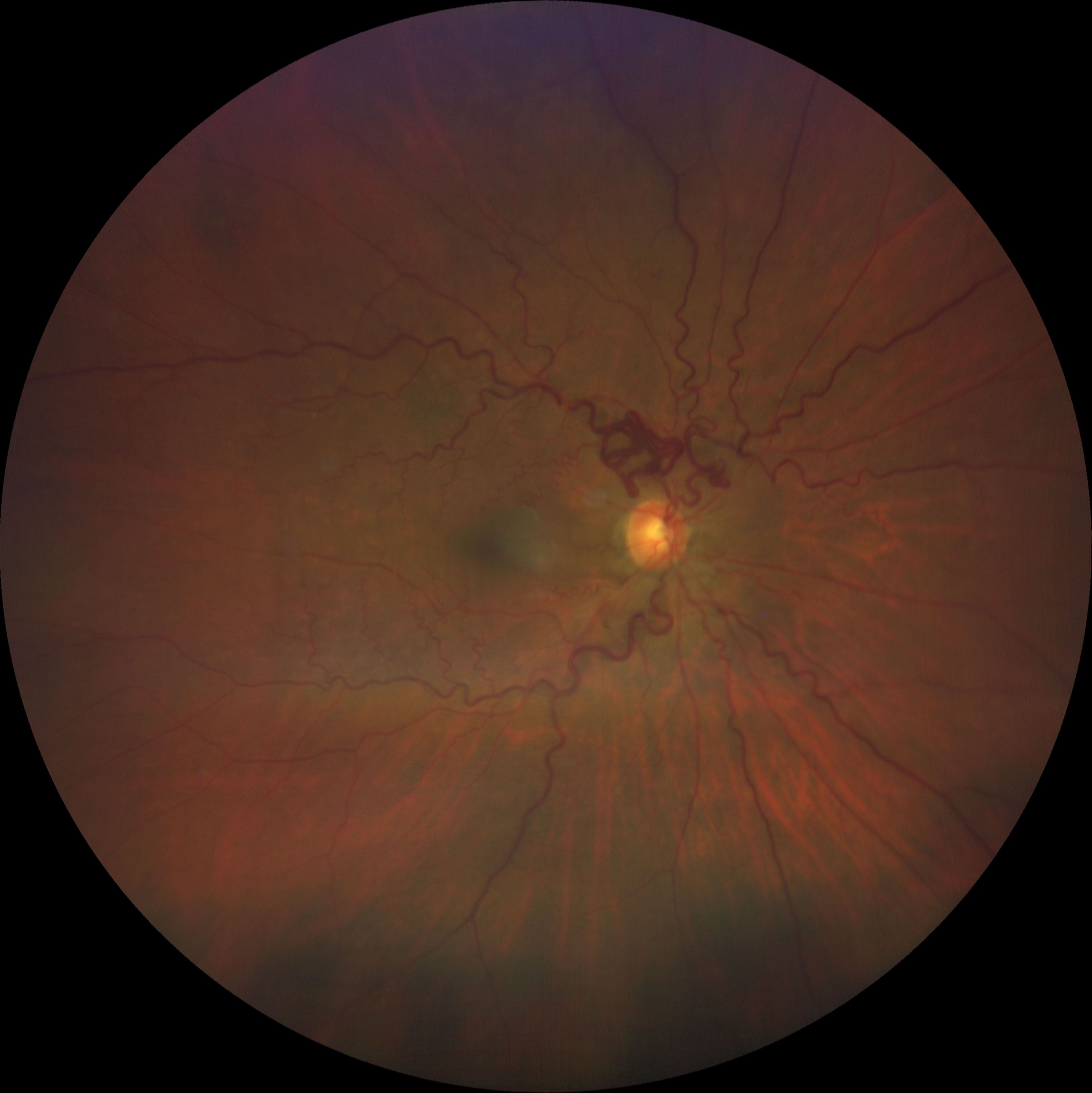
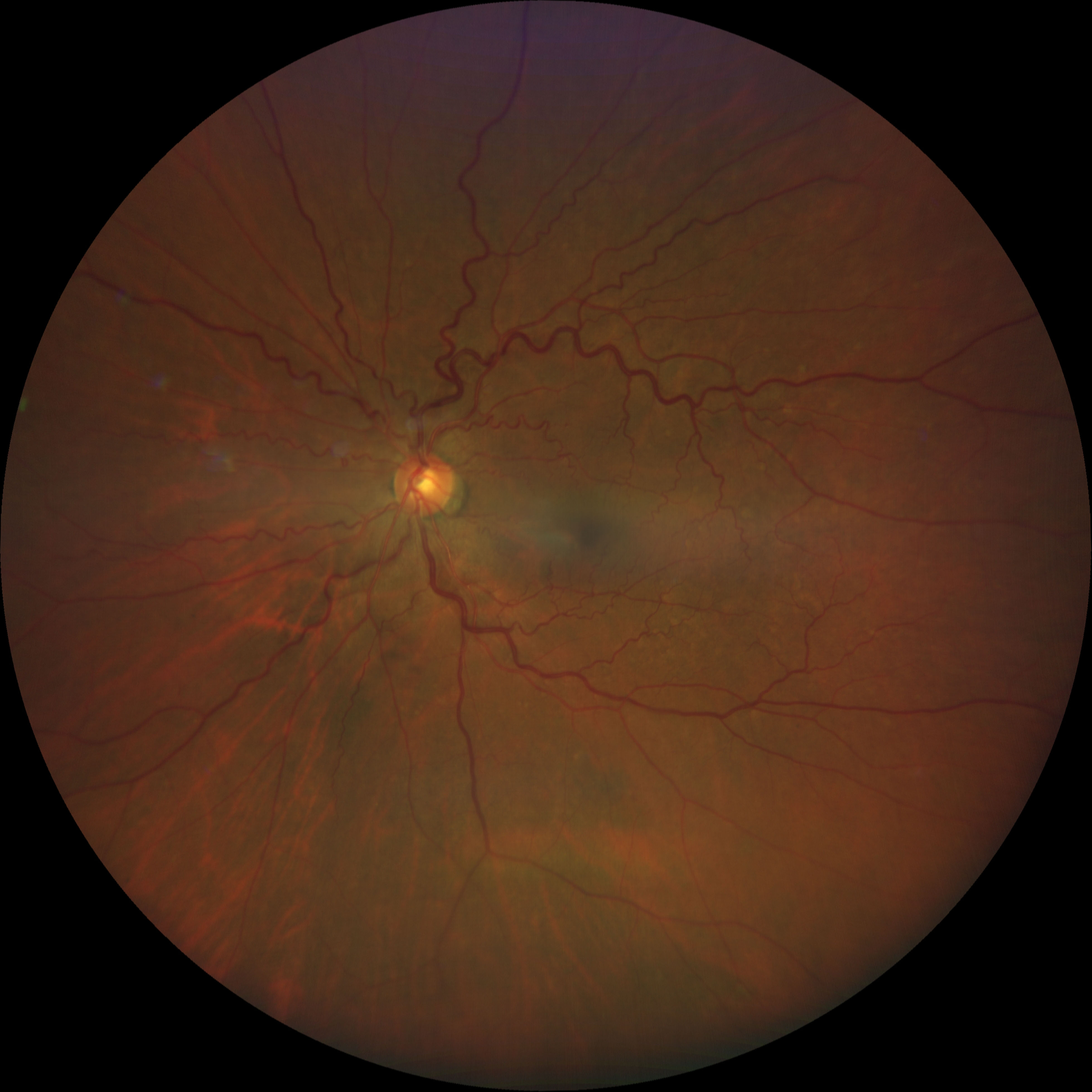
A 64-year-old woman was found on routine examination to have severe tortuosity of the superior arcade vessels in the right eye consistent with an arterial venous malformation (AVM). The other eye had moderately dilated and tortuous retinal vasculature. Retinal AVMs can be seen in isolation as an acquired anomaly or as an ophthalmic finding in Wyburn-Mason Syndrome, a rare congenital disorder characterized by AVMs in the retina and brain. Vision loss in AVMs may occur due to complications such as vascular occlusion. In this patient, MRI/MRA brain was negative for brain AVMs, and regular surveillance was advised.

Ophthalmic Atlas Images by EyeRounds.org, The University of Iowa are licensed under a Creative Commons Attribution-NonCommercial-NoDerivs 3.0 Unported License.


Address
University of IowaLegal
Related Links