This site uses tracking information. Visit our privacy policy. Click to agree to this policy and not see this again.

Ophthalmology and Visual Sciences

Rhegmatogenous Retinal Detachment, Macula-Off

Rhegmatogenous Retinal Detachment, Macula-Off

Category(ies): Retina, Vitreous
Contributor: Eric Chin, MD
Photographer: Cindy Montague, CRA

Patient

  • • A 62 year old female with a history of uncomplicated cataract surgery in both eyes, presented to the comprehensive clinic for decreased vision in the left eye since one day prior. She said her nasal field of vision had become abruptly and increasingly blurry while doing the laundry. She denied any flashes, floaters, or other complaints. She denied prior trauma or recent falls. She had no family history of retinal detachments.
  • • Vision: OD 20/25-3; OS 20/100+1
  • • IOP: 14 and 13
  • • Manifest refraction prior to cataract surgery:
    • º OD: -13.25 +0.75 045
    • º OS: -9.25 +0.50 040
    • º Slit lamp exam: Posterior chamber intraocular lenses and posterior capsular opacities, both eyes
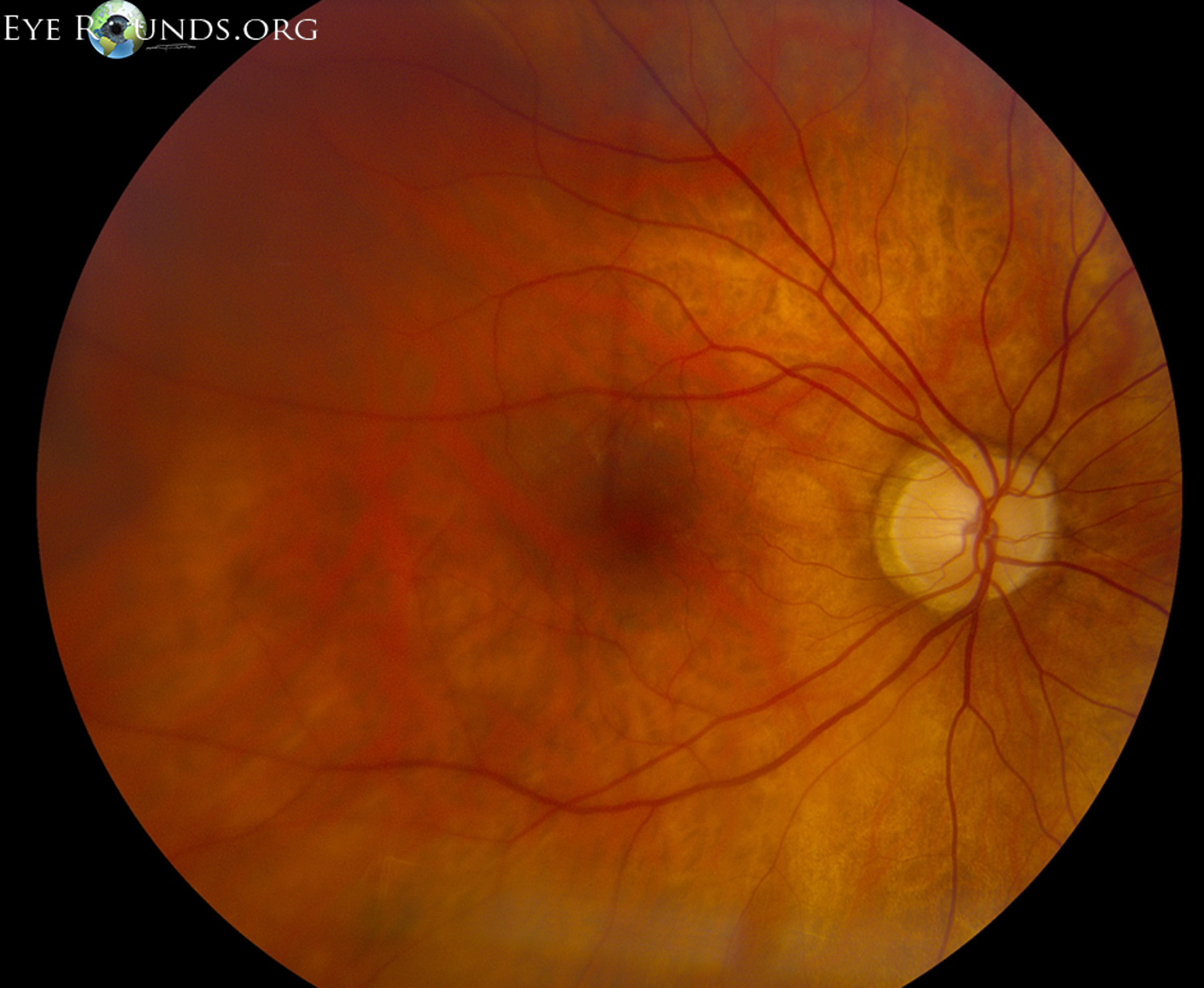
fundus photo
Image 1: OD: clear media; normal disc, vessels and macula; in the periphery, there were scattered areas of lattice degeneration (270 degrees from 4 to 1 o'clock clockwise) near the ora, however there were no other retinal tears or detachments.
OD OCT
Image 2: OD OCT: normal
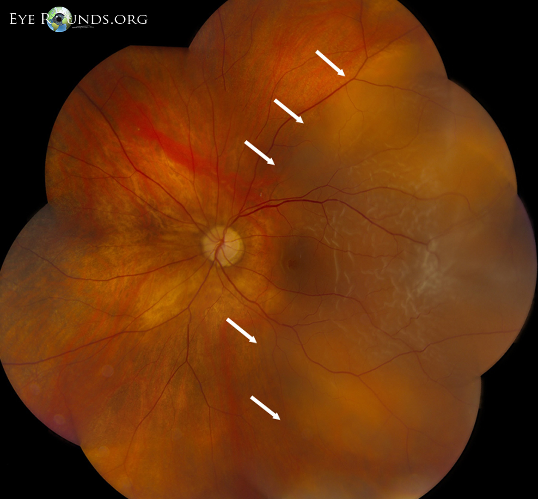
rhegmatogenous macula-off retinal detachment
Image 3: OS: low magnification montage, rhegmatogenous macula-off retinal detachment (temporal to the white arrows); not shown, the patient had a horseshoe retinal tear at 1 o'clock near the ora, as well as multiple areas of lattice degeneration near the ora from 10 to 2 o'clock and 4 to 8 o'clock clockwise. Intraoperatively, there were multiple holes seen in her lattice degeneration at 5 o'clock and 6:30 o'clock.
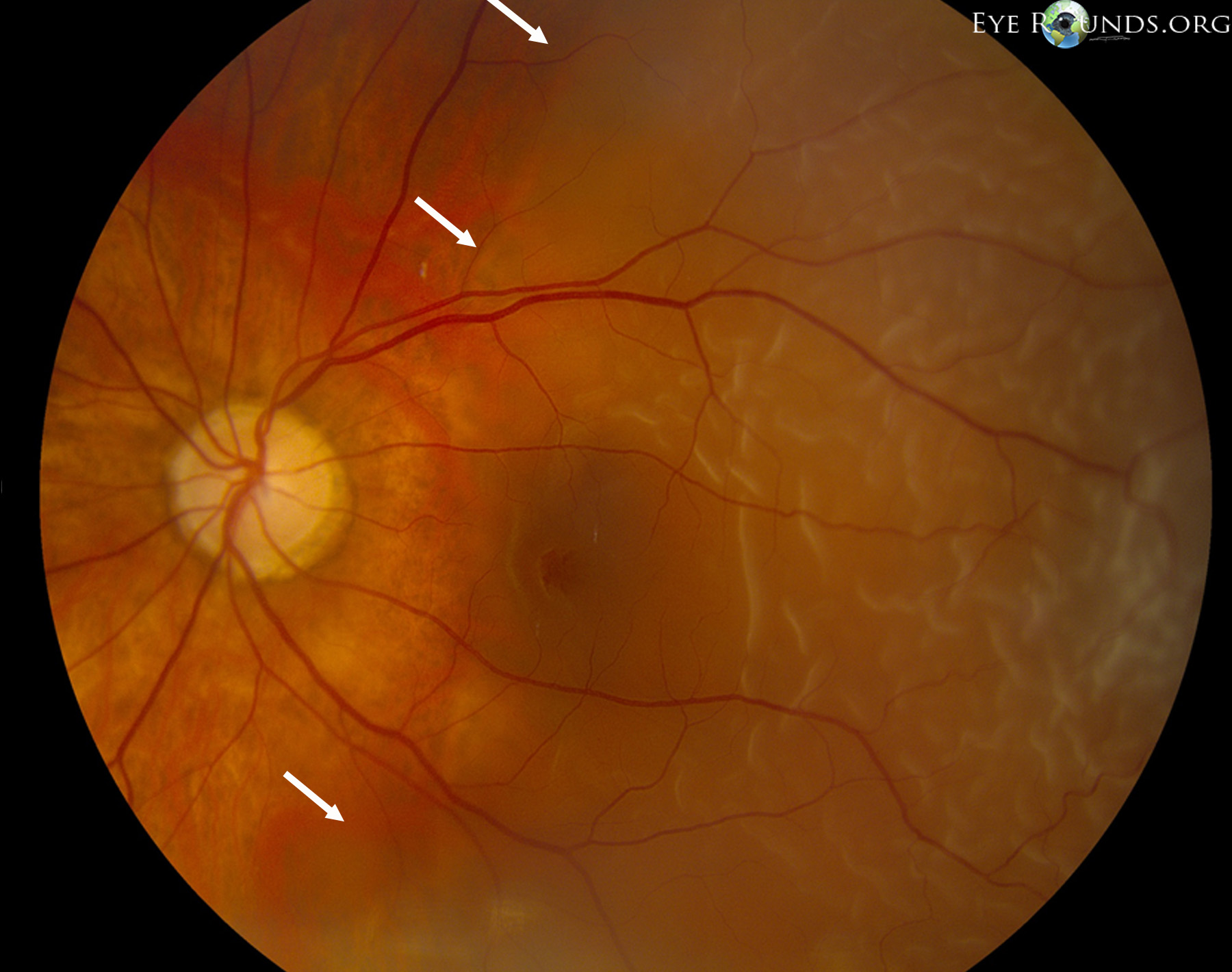
higher magnification of the rhegmatogenous retinal detachment
Image 4: OD: higher magnification of the rhegmatogenous retinal detachment that involves the macula.
oct limited quality, macula-off retinal detachment without a macular hole
Image 5: OCT OS: limited quality, macula-off retinal detachment without a macular hole.
  • The patient underwent a scleral buckle, 25-gauge pars plana vitrectomy, laser indirect ophthalmoscopy, air-fluid exchange, and injection of 22% SF6, in the left eye.
  • The right eye was also treated with prophylactic laser indirect ophthalmoscopy intraoperatively.

Risk Factors for Retinal Detachments

  • Age*
  • High myopia*
  • Co-morbid eye conditions, such as lattice degeneration*
  • Prior eye surgery*
  • Prior retinal detachment
  • Family history of retinal detachment
  • Trauma

*Risk factors seen in our patient are highlighted in bold.