The patient was a 57-year-old man with vision loss in the right eye (OD) greater than left eye (OS) in the setting of a craniopharyngioma, which was resected 16 years prior. His visual acuity had been no light perception OD and 20/20 OS since the procedure.
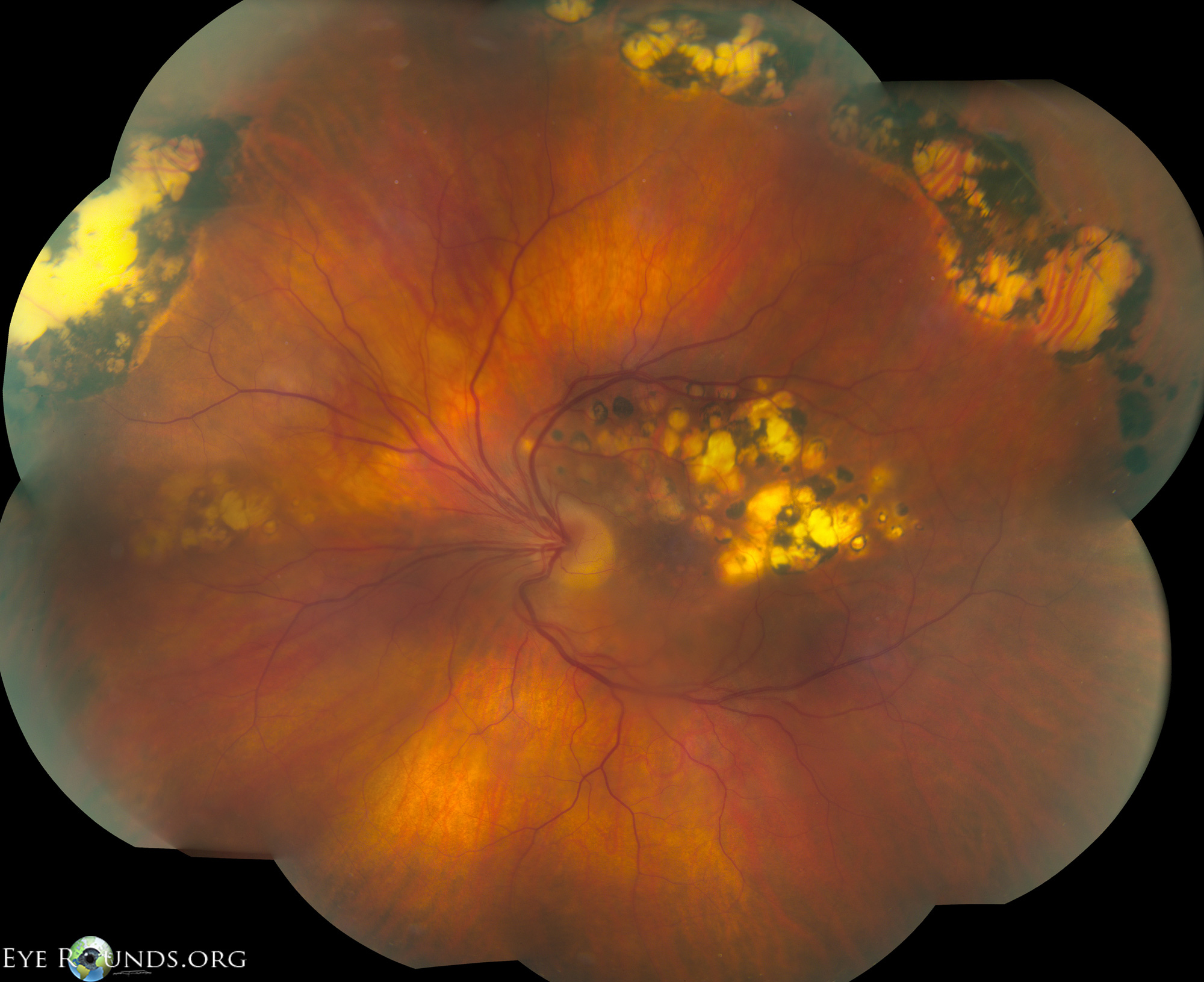
Figure 1 is a fundus photo from a patient who has significant situs inversus (note, the scarring in the macula is from prior laser treatment and unrelated to the situs inversus).
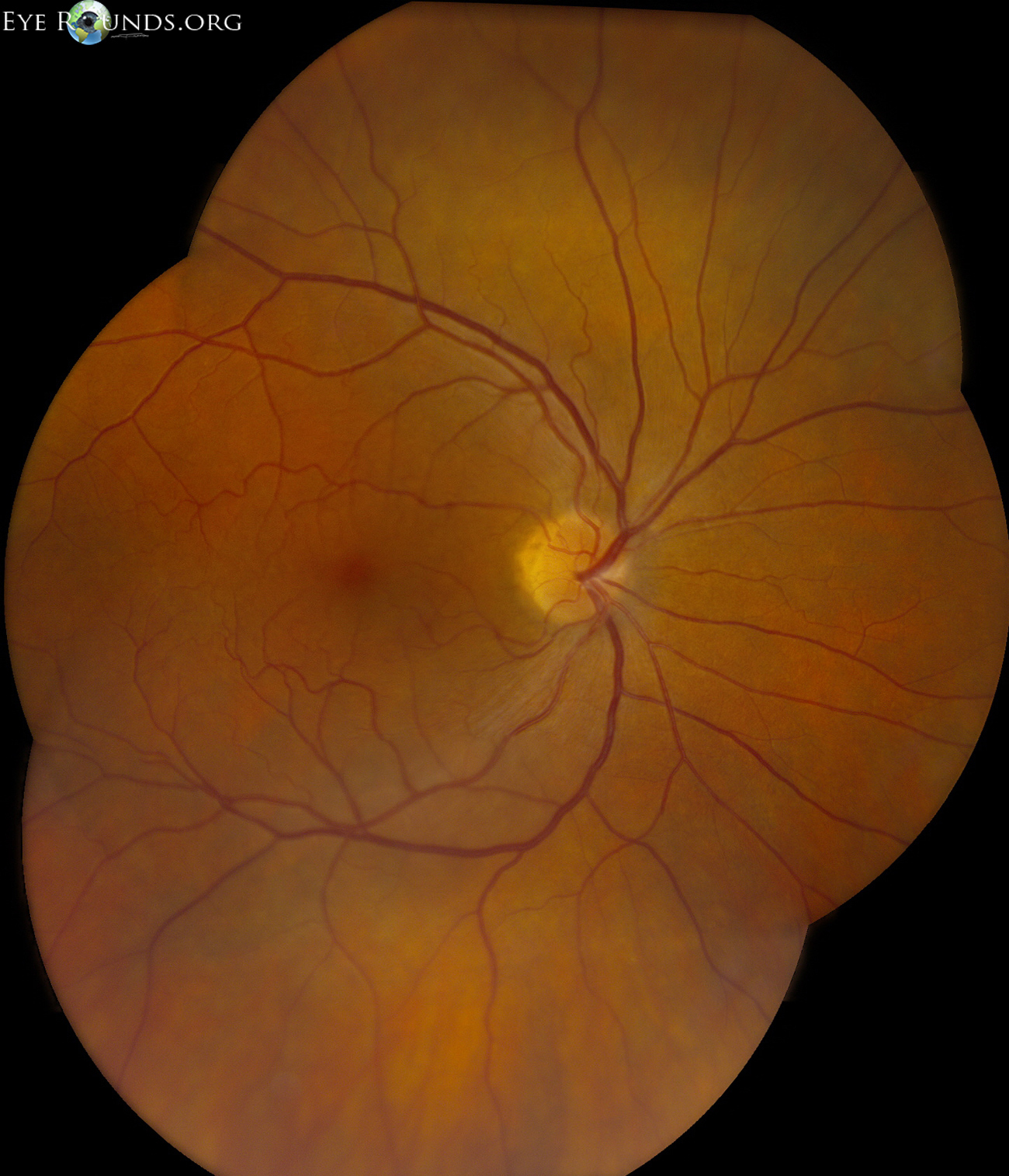
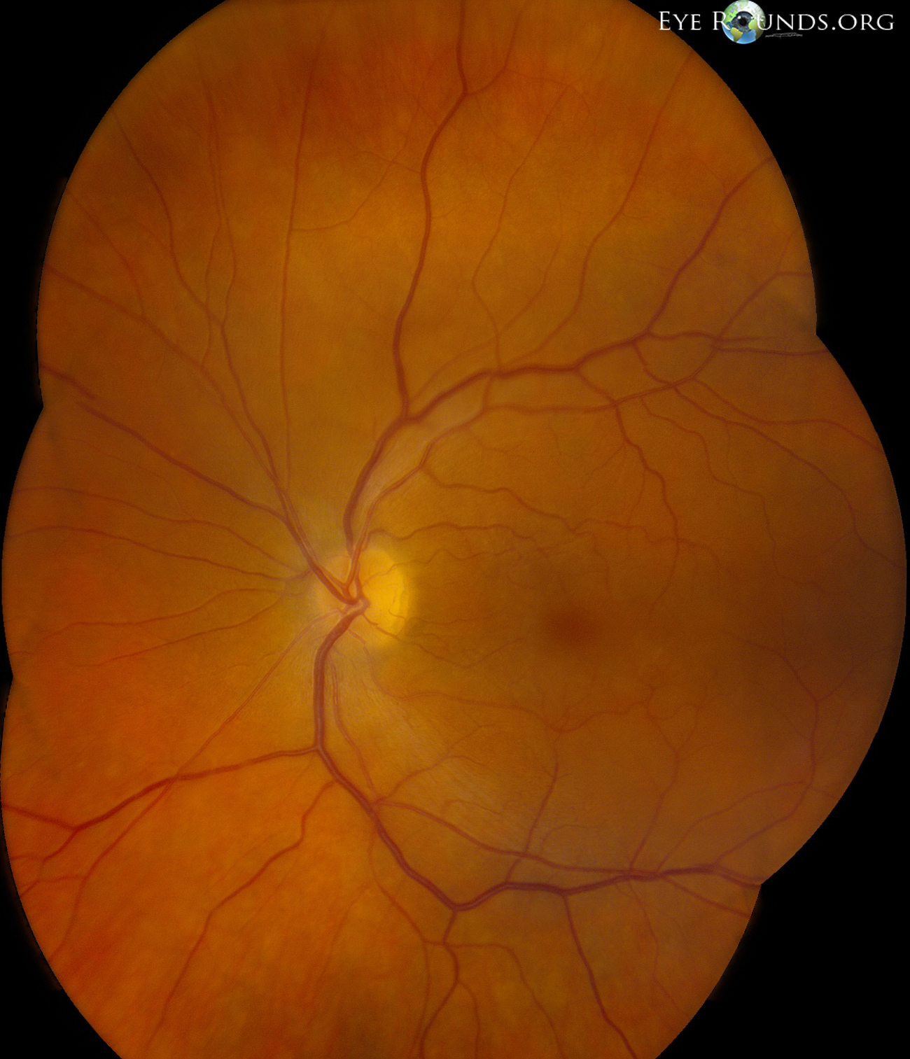
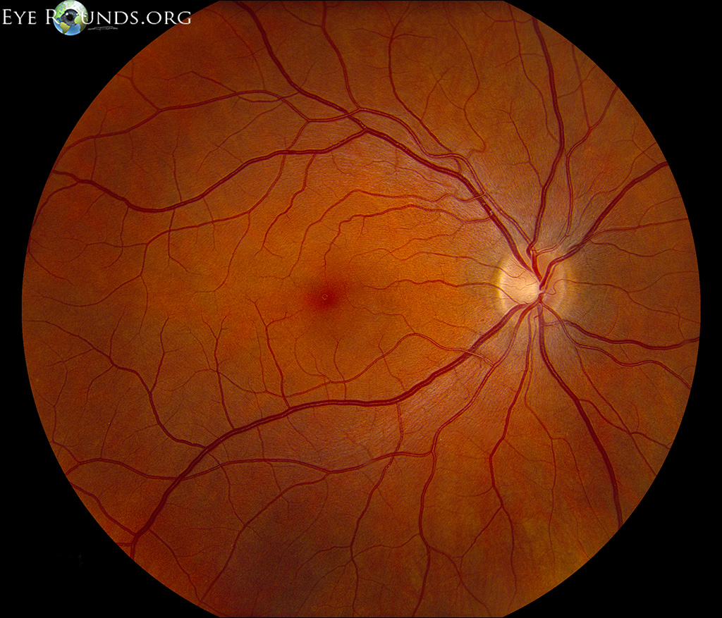
Figure 2 is a fundus photo from a different patient who has moderate situs inversus. It is easiest to appreciate this vascular variation when it is compared to a normal fundus (Figure 3).

Ophthalmic Atlas Images by EyeRounds.org, The University of Iowa are licensed under a Creative Commons Attribution-NonCommercial-NoDerivs 3.0 Unported License.


Address
University of IowaLegal
Related Links