This site uses tracking information. Visit our privacy policy. Click to agree to this policy and not see this again.

Ophthalmology and Visual Sciences

The Sympathizing Eye: Panuveitis Secondary to Sympathetic Ophthalmia

The Sympathizing Eye: Panuveitis Secondary to Sympathetic Ophthalmia

Category(ies): Retina, Uveitis
Contributor: Brittni A. Scruggs, MD, PhD and Ian C. Han, MD

This is a 46-year-old monocular man who presented with floaters, peripheral vision loss, and decreased central acuity in his left eye. Nine months prior to presentation, the right eye had suffered a penetrating globe injury with retinal detachment and was enucleated within four weeks of the injury.

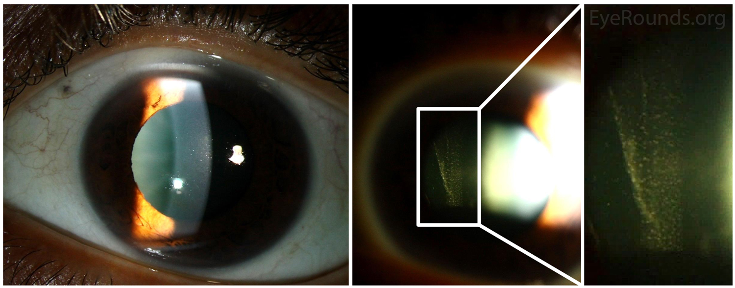
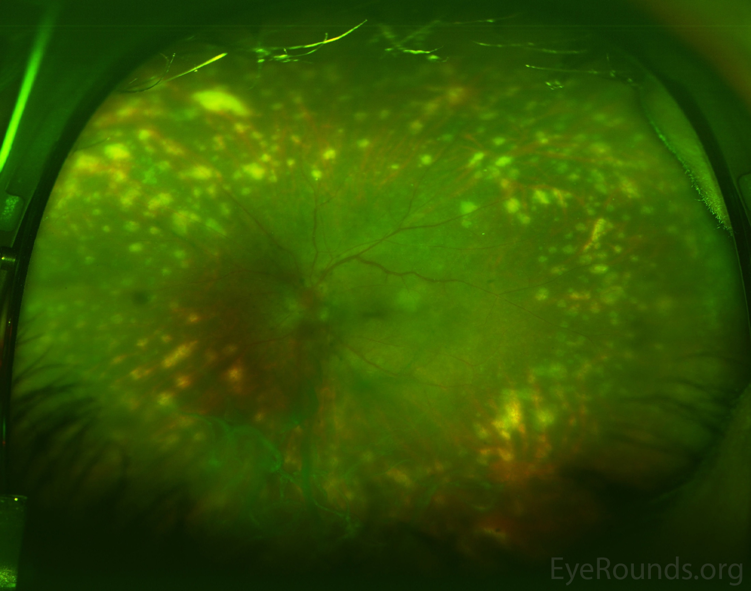
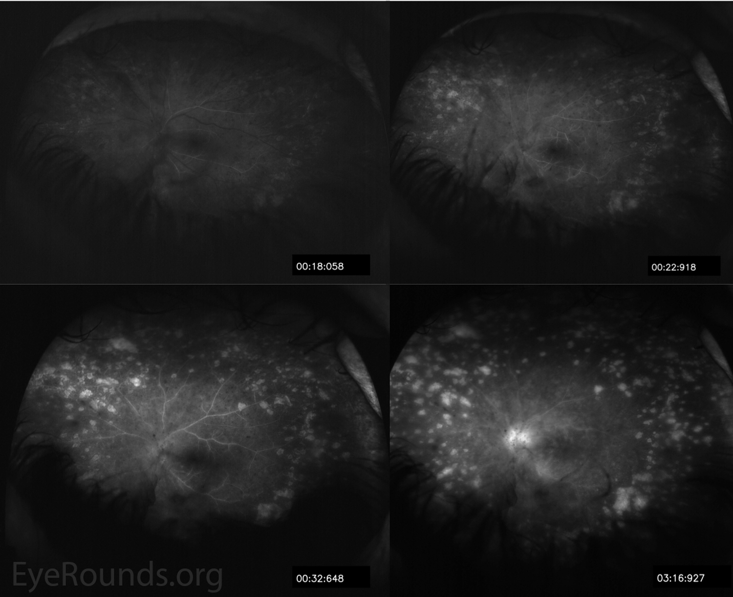
His slit lamp exam demonstrated anterior and posterior chamber cell (Figure 1). Dilated fundus exam revealed vitreous strands, grade III disc edema, and multiple, discrete, peripheral hypopigmented lesions consistent with Dalen-Fuchs nodules (i.e., lymphocytes and epithelioid histiocytes present between the RPE and Bruch membrane) (Figure 2). Fluorescein angiography demonstrated staining of the multiple peripheral lesions and leakage from the optic nerve and vessels in the posterior pole (Figure 3).

Despite initial treatment with mycophenolate mofetil (CellCept) for several months, the patient experienced worsening peripheral vision and persistent vitreous inflammation. He underwent surgical placement of a fluocinolone acetonide intravitreal implant (Retisert) for local, long-term inflammation control.

Sympathetic ophthalmia (SO) is a rare, granulomatous panuveitis that usually occurs within one year of penetrating trauma to the fellow eye, likely as a result of uveoretinal antigens entering the conjunctival lymphatics [1, 2]. In addition to trauma, pars plana vitrectomy and other intraocular procedures are associated with increased SO risk [2]. Other characteristic findings of SO that were not present in this case include serous retinal detachments and mutton-fat keratic precipitates.

Please refer to the Eye Rounds case on sympathetic ophthalmia for further information regarding work-up, differential diagnosis, pathophysiology, and management.

 Slitlamp photography
Figure 1. Slitlamp photography, left eye. There was 1+ cell and 1+ flare in the anterior chamber with trace nuclear sclerosis (left image). The vitreous exam was remarkable for significant anterior vitritis (right images).
 Optos ultra-widefield pseudocolor fundus photography
Figure 2. Optos ultra-widefield pseudocolor fundus photography, left eye. There were numerous hypopigmented, atrophic-appearing chorioretinal lesions throughout the mid- and far- periphery that were consistent with Dalen-Fuchs nodules. There was grade III disc edema, venous engorgement, and peripheral vascular attenuation.
 Optos ultra-widefield pseudocolor fundus photography
Figure 3. Fundus fluorescein angiography, left eye. Irregular filling of the choroid was present. There was leakage from the optic nerve and vessels throughout the posterior pole. Hyperfluorescence of atrophic areas was seen.
 Optos ultra-widefield pseudocolor fundus photography
Figure 4. Optical coherence tomography of the macula, left eye (OS). There was a moderate epiretinal membrane with mild intraretinal fluid and loss of the foveal contour OS. There was marked disc edema with fluid tracking into the nasal macula OS; however, no subretinal fluid was present subfoveally.
References:
  1. Aziz HA, Flynn HW, Young RC, Davis JL, Dubovy SR. SYMPATHETIC OPHTHALMIA: Clinicopathologic Correlation in a Consecutive Case Series. Retina 2015;35(8):1696-1703. https://PubMed.gov/25719985. DOI: 10.1097/IAE.0000000000000506
  2. Tan XL, Seen S, Dutta Majumder P, Ganesh SK, Agarwal M, Soni A, Biswas J, Aggarwal K, Mahendradas P, Gupta V, Ling HS, Teoh S, Pavesio C, Agrawal R. Analysis of 130 Cases of Sympathetic Ophthalmia - A Retrospective Multicenter Case Series. Ocul Immunol Inflamm 2018;10.1080/09273948.2018.1517894:1-8. https://PubMed.gov/30207811. DOI: 10.1080/09273948.2018.1517894

Image Permissions:
Creative Commons

Ophthalmic Atlas Images by EyeRounds.org, The University of Iowa are licensed under a Creative Commons Attribution-NonCommercial-NoDerivs 3.0 Unported License.