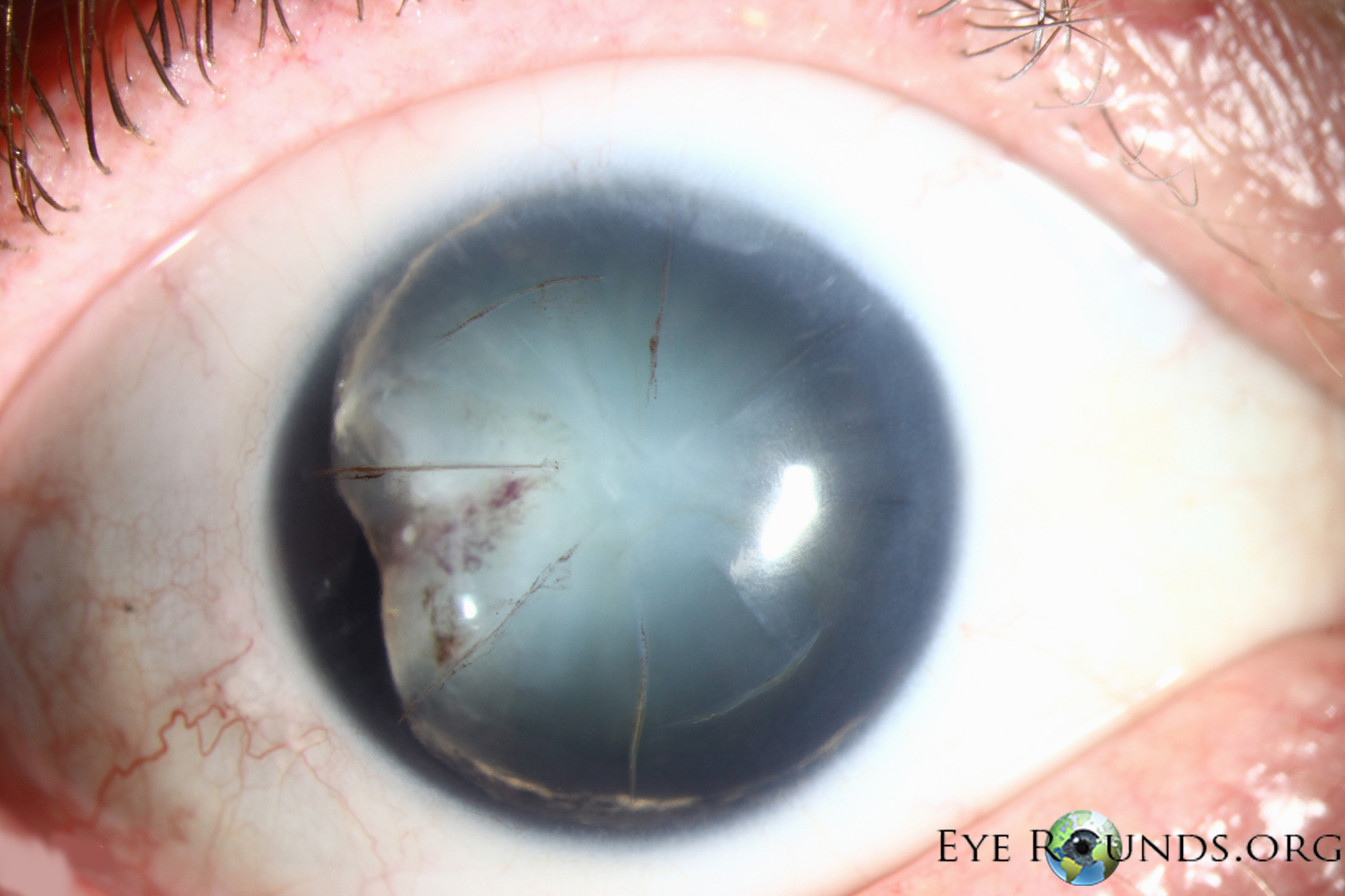
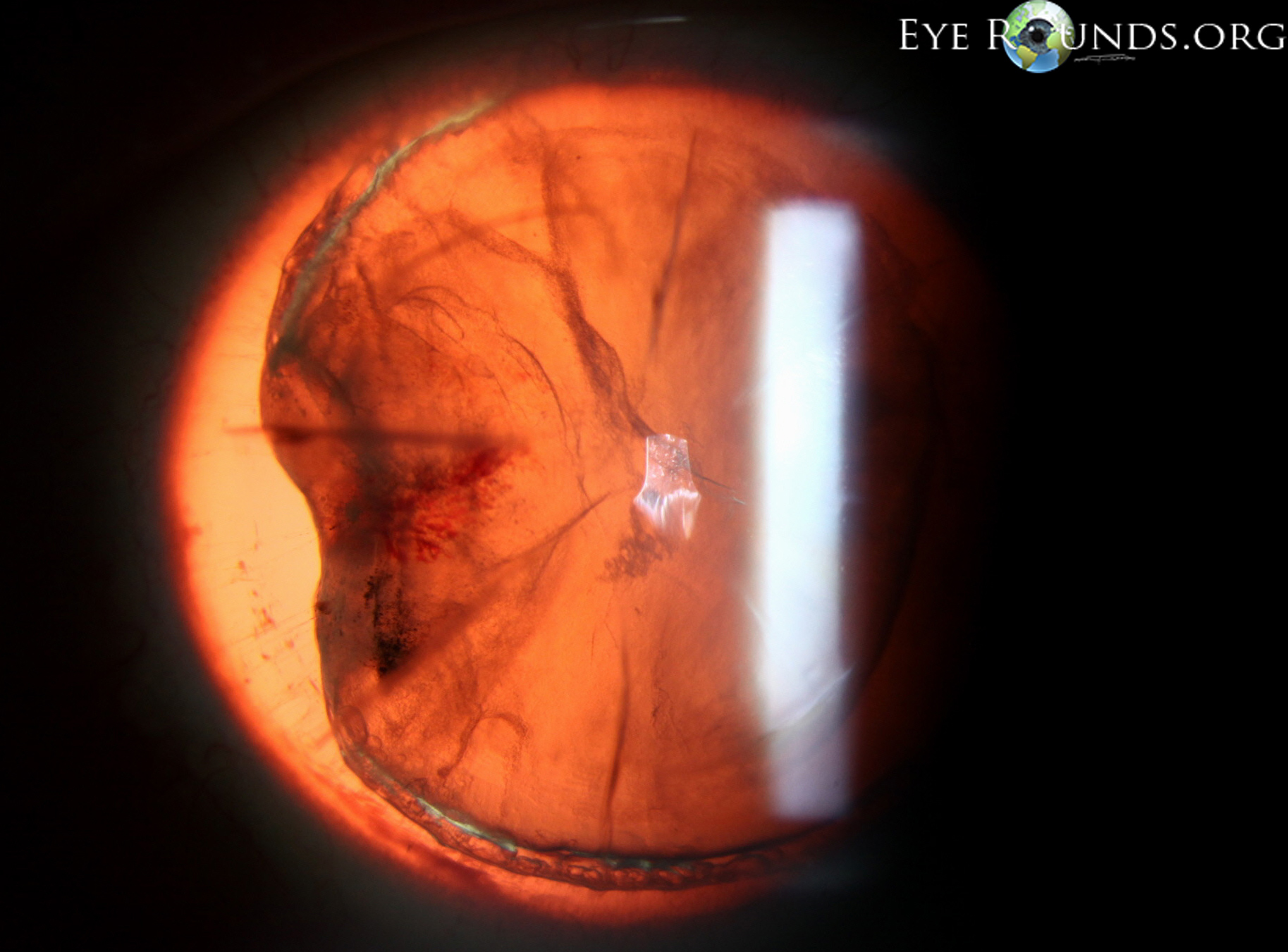
59-year-old male with past ocular history of radial keratotomy (RK) in both eyes, presented to our clinic after sustaining an injury to his right eye with a wrench. Initially there was nearly a complete hyphema and no view of iris, lens, or posterior pole. B scan echography did not show any posterior pathology. Several months later, after the hyphema had resolved, the patient returned with the exam seen below. His vision was hand motions at this time and he was noted to have a traumatic cataract in the right eye. Interestingly, the iris was noted to be missing on exam. There was no evidence of even a small iris stump on gonioscopy. Previous photos of the patient confirmed that he did, at one time, have both a clear lens and complete iris.
He was also noted to have pigment on his RK scars. After discussion amongst several faculty, the current hypothesis is that he may have had an open globe with the initial injury, and the iris may have prolapsed through one of his RK scars. This may have been inadvertently wiped from the eye by the patient, prior to his seeking medical care. The RK wound may have then self-sealed.
This patient was referred to another ophthalmologist for cataract extraction and insertion of a pupillary prosthesis.

Ophthalmic Atlas Images by EyeRounds.org, The University of Iowa are licensed under a Creative Commons Attribution-NonCommercial-NoDerivs 3.0 Unported License.


Address
University of IowaLegal
Related Links