

EyeRounds Online Atlas of Ophthalmology
Contributor: William Charles Caccamise, Sr, MD, Retired Clinical Assistant Professor of Ophthalmology, University of Rochester School of Medicine and Dentistry
*Dr. Caccamise has very generously shared his images of patients taken while operating during the "eye season" in rural India as well as those from his private practice during the 1960's and 1970's. Many of his images are significant for their historical perspective and for techniques and conditions seen in settings in undeveloped areas.
Category: Trauma
Traumatic iridodialysis with slight anterior cortical traumatic cataract
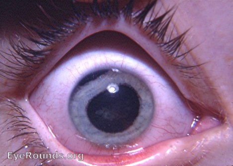
The telltale characteristic of an iridodialysis is present in this photo, i.e. the flattening of the pupil margin curve in that part of the iris that has been torn from its ciliary body attachment.A chord effect results in that part of the pupil margin. Of course, the empty area between the torn edge of the iris and the limbus is the site of the iridodialysis. Gonioscopy will give a full view of the detachment site. There is also a nebular anterior cortical traumatic cataract.

Ophthalmic Atlas Images by EyeRounds.org, The University of Iowa are licensed under a Creative Commons Attribution-NonCommercial-NoDerivs 3.0 Unported License.