

EyeRounds Online Atlas of Ophthalmology
Contributor: William Charles Caccamise, Sr, MD, Retired Clinical Assistant Professor of Ophthalmology, University of Rochester School of Medicine and Dentistry
*Dr. Caccamise has very generously shared his images of patients taken while operating during the "eye season" in rural India as well as those from his private practice during the 1960's and 1970's. Many of his images are significant for their historical perspective and for techniques and conditions seen in settings in undeveloped areas.
Category: Systemic Disorders
Tropical eosinophilia with bulbar conjunctival hyperemia, both eyes
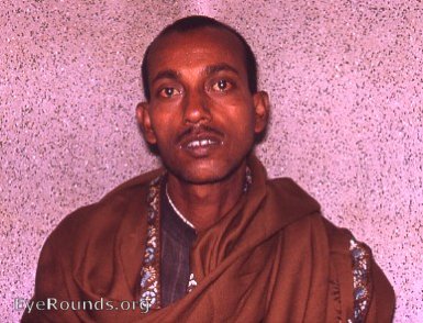
The Kurji Holy FamilyHospital Eye Clinic was located in an area highly endemic for microfilariasis. Those patients who had unexplained bulbar conjunctival hyperemia were screened for tropical eosinophilia by blood counts for eosinophilia, by chest x-ray examination for lung infiltrates, and for blood smears for the remote possibility of finding microfilariae.Through these studies, it was possible to make a diagnosis of tropical eosinophilia in this young man. He responded to treatment for microfilariasis.
Infection with the Wucheria bancrofti is endemic in the Eye Clinic patient-population. The microfilariae are transmitted from the heavy mosquito population to the heavy easily bitten human population of the area. When a mosquito sucks blood from an already infected person, it withdraws with the blood a number of filaria embryo. These then undergo stages of development in the body of the mosquito. Finally, these larvae (microfilariae) reach the proboscis of the mosquito and are injected into the next victim of that mosquito's unfriendly bite.
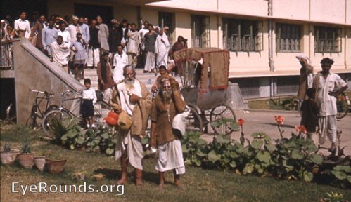
The sadhu with the orange bucket has a markedly swollen stove-pipe left leg due to his chronic filariasis- lymphedema of the left leg.

Ophthalmic Atlas Images by EyeRounds.org, The University of Iowa are licensed under a Creative Commons Attribution-NonCommercial-NoDerivs 3.0 Unported License.