This site uses tracking information. Visit our privacy policy. Click to agree to this policy and not see this again.

Ophthalmology and Visual Sciences

Xanthelasma

Xanthelasma

Category(ies): Cornea, External Eye Disease, Oculoplastics
Contributor: Austin R. Fox, MD, Angela Mahoney, and Audrey C. Ko, MD
Photographer: Audrey C. Ko, MD

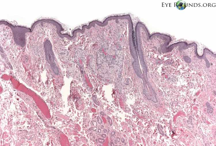
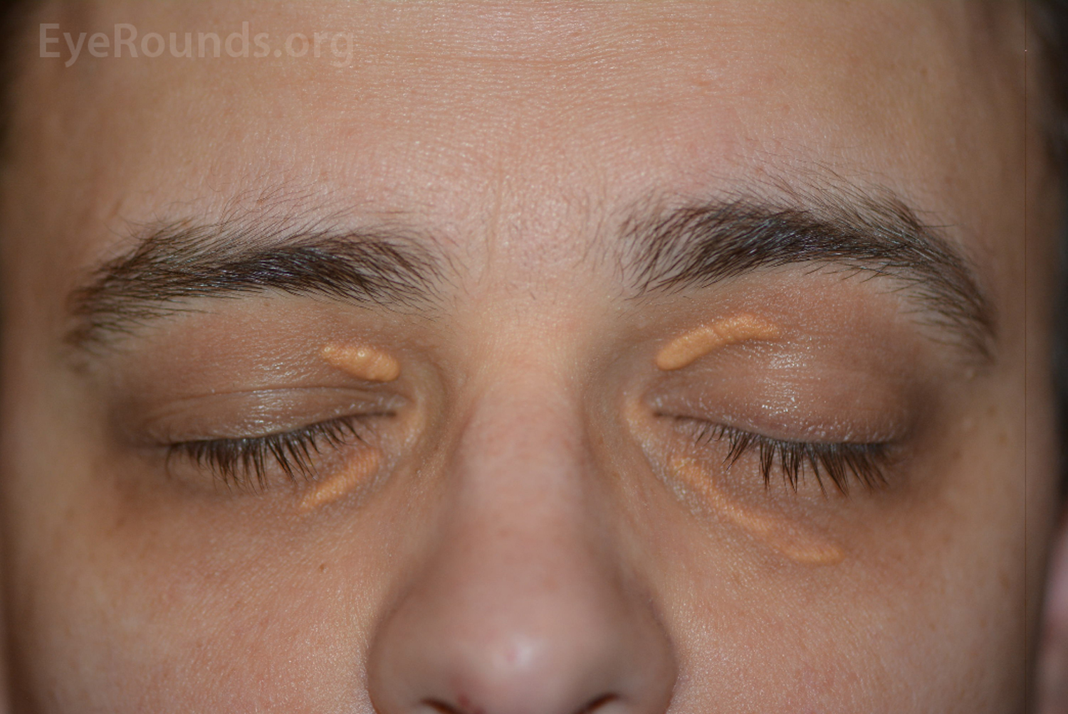
Xanthelasmas are yellowish papules or plaques caused by the deposition of lipid on or around the eyelids. The lesions are relatively rare, with an incidence of 1.1% in women and 0.3% in men [1]. Lesions typically appear between 40 and 60 years but may appear at younger ages as a sign of a familial dyslipoproteinemia. Approximately 50% of patients with xanthelasmas have abnormal cholesterol or triglyceride levels, and these lesions have been associated with hypothyroidism, cirrhosis, and nephrotic syndrome. Therefore, systemic work-up is indicated in these patients. Treatment of these lesions include observation, surgical excision, topical trichloroacetic acid (TCA), laser ablation, and liquid nitrogen cryotherapy [2]. Regardless of the treatment modality, recurrence is common and is reported to be 40% after primary surgical excision [3].

middle-aged man presented with bilateral symmetric yellowish lesions on the medial upper and lower eyelids
Figure 1. A healthy middle-aged man presented with bilateral symmetric yellowish lesions on the medial upper and lower eyelids. These lesions had slowly enlarged over a three year period. His past medical history was unremarkable for hyperlipidemia, diabetes mellitus, obesity, or other systemic disease. In this case, the patient opted for surgical excision of these lesions due to cosmetic concerns.
Reference:
  1. Rohrich, R.J., J.E. Janis, and P.H. Pownell, Xanthelasma palpebrarum: a review and current management principles. Plast Reconstr Surg, 2002. 110(5): p. 1310-4.
  2. Laftah, Z. and F. Al-Niaimi, Xanthelasma: An Update on Treatment Modalities. J Cutan Aesthet Surg, 2018. 11(1): p. 1-6.
  3. Mendelson, B.C. and J.K. Masson, Xanthelasma: follow-up on results after surgical excision. Plast Reconstr Surg, 1976. 58(5): p. 535-8.

Image Permissions:
Creative Commons

Ophthalmic Atlas Images by EyeRounds.org, The University of Iowa are licensed under a Creative Commons Attribution-NonCommercial-NoDerivs 3.0 Unported License.