This site uses tracking information. Visit our privacy policy. Click to agree to this policy and not see this again.

Ophthalmology and Visual Sciences

Granular Corneal Dystrophy Discovered Following LASIK:

A Clinicopathologic Correlation of Granular Corneal Dystrophy Type II and LASIK

Granular Corneal Dystrophy Discovered Following LASIK:

A Clinicopathologic Correlation of Granular Corneal Dystrophy Type II and LASIK
Alexander S. Davis, PhD and Nasreen Syed, MD
September 16, 2011

Chief Complaint: Decreased vision

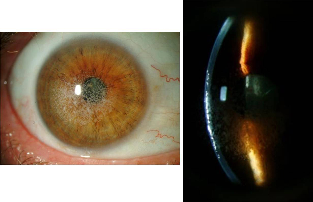
History of Present Illness: A 44-year-old male presented to the University of Iowa Hospitals and Clinics for progressive decrease in vision in both eyes (OU) with "tremendous" glare symptoms.

Past Ocular History: Laser-assisted in situ keratomileusis (LASIK) OU in 2002 (8 years prior to presentation)

Past Medical History: Non-contributory

Medications: Clozapine, Ibuprofen, lithium, multivitamin

Allergies: None

Family History: Brother has similar corneal problems without LASIK. Mother had cataracts.

Social History: No alcohol use but smokes tobacco

OCULAR EXAMINATION

  • Visual Acuity (without correction):
    • OD: 20/25 -1 (no improvement with pinhole)
    • OS: 20/40 -1 → 20/30 -1 (with pinhole)
  • Confrontation Visual Fields: Full OU
  • Motility: Full OU
  • Slit Lamp Exam: (examination descriptions are OU unless specified)
    • Lids/Lashes: Normal
    • Conjunctiva/Sclera: Clear and quiet
    • Cornea: LASIK flap; granular deposits in interface
    • Anterior Chamber: Deep and quiet
    • Iris: Normal architecture
    • Lens: 1+ nuclear sclerotic cataract
  • Figure 1. Slit lamp photos of right eye demonstrating granular white opacities located at the level of the LASIK flap interface (click image for higher resolution photo)
    Slit lamp photos of right eye showing granular white opacities at the LASIK flap interface level

    Figure 2. Slit lamp photos of left eye demonstrating granular white opacities located at the level of the LASIK flap interface (click image for higher resolution photo)
    Slit lamp photos of left eye demonstrating granular white opacities located at the level of the LASIK flap interface

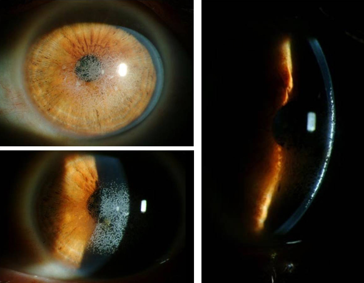
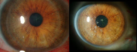
Clinical Course: Following the initial visit, the patient underwent a superficial keratectomy followed by phototherapeutic keratectomy (PTK) OU without complications. A slit lamp exam on the first post-operative day showed a reduction in the density of granular deposits in the visual axis. On post-operative day 7 the patient's vision remained poor at all distances, with visual acuity of 20/80 -1 OU. Due to the patient's poor vision, Intralase-enabled superficial anterior lamellar keratoplasty (IE-SALK) was recommended for both eyes, starting with the right eye. Slit-lamp photos of both eyes were obtained prior to surgery and are shown in figures 3 and 4.

Figure 3. Slit lamp photos of the right eye, following superficial keratectomy and phototherapeutic keratectomy (PTK), but prior to Intralase-enabled superficial anterior lamellar keratoplasty (click image for higher resolution photo)
Slit lamp photos of the right eye, following superficial keratectomy and phototherapeutic keratectomy (PTK), but prior to Intralase-enabled superficial anterior lamellar keratoplasty

Figure 4. Slit lamp photos of the left eye after superficial keratectomy and PTK (click image for higher resolution photo)
Slit lamp photos of the left eye after superficial keratectomy and PTK

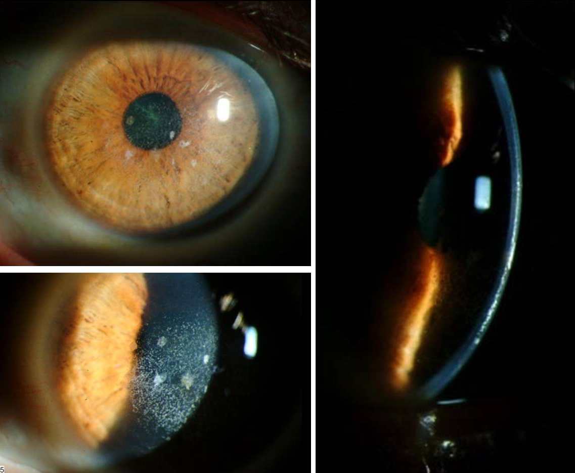
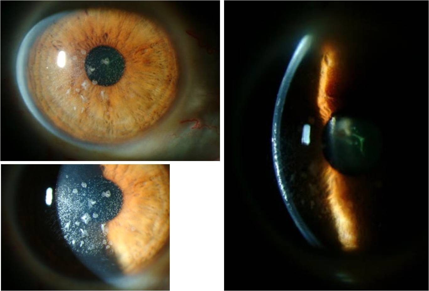
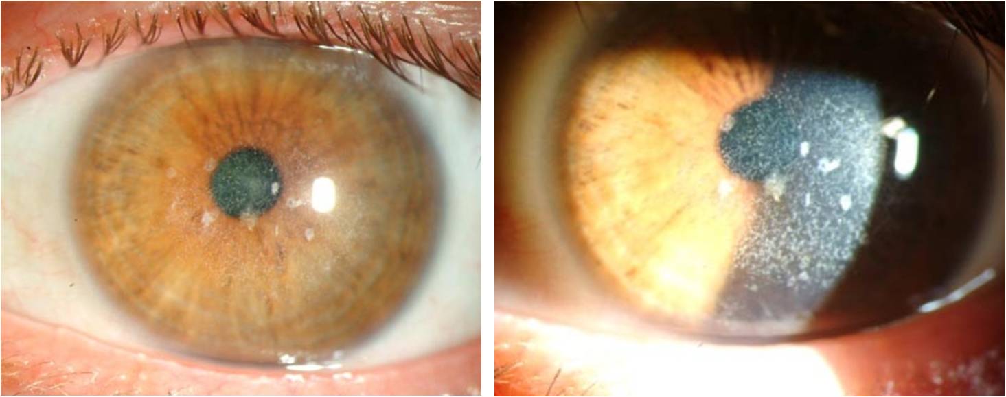
On post-operative day 3 after IE-SALK of the right eye, vision had not improved OD. However, although IE-SALK was not performed on the fellow eye, visual acuity had improved to 20/40 without correction OS since the superficial keratectomy and PTK performed over a month ago.(see figures 5 and 6 for photos of OD and OS, respectively).

Figure 5. Slit lamp photo of the right eye POD#3 after IntraLase-enabled superficial anterior lamellar keratoplasty, with preoperative photo on the right for comparison (click image for higher resolution photo)
Slit lamp photo of the right eye POD#3 after IntraLase-enabled superficial anterior lamellar keratoplasty, with preoperative photo on the right for comparison (click image for higher resolution photo)

Figure 6. Slit lamp photo of left eye, after superficial keratectomy and PTK (>30 days ago), taken at the time of OD post-operative photo (Figure 5), without significant worsening since initial treatment (click image for higher resolution photo)
Slit lamp photo of left eye, after superficial keratectomy and PTK (>30 days ago), taken at the time of OD post-operative photo (Figure 5), without significant worsening since initial treatment (click image for higher resolution photo)

Post-operative Pathology

  • Specimen: LASIK flap from right eye
  • Stains used: Hematoxylin and eosin (H&E), Periodic acid Schiff (PAS), Masson trichrome, Congo red
Figure 7. Eosinophilic deposits are noted in the anterior stroma and at the stromal interface (H&E, original magnification= 50X) (click image for higher resolution photo)
Eosinophilic deposits are noted in the anterior stroma and at the stromal interface

Figure 8. Eosinophilic deposits stain bright red with Masson trichrome stain (original magnification = 50X with 200X insert) (click image for higher resolution photo)
Eosinophilic deposits stain bright red with Masson trichrome stain

Figure 9. Stromal deposits do not stain with Congo red stain or demonstrate birefringence with cross-polarization (original magnification = 100X) (click image for higher resolution photo)
Stromal deposits do not stain with Congo red stain or demonstrate birefringence with cross-polarization

Pathological Diagnosis: Granular Corneal Dystrophy

DISCUSSION

In general, all corneal dystrophies are classified by the anatomical layer of the cornea that is involved. This has been the case even after re-classification of the dystrophies in recent years (Weiss 2008, Birkholz 2009; please see the excellent review by Birkholz et al. for a more detailed review of corneal dystrophies: http://www.eyerounds.org/cases/43-Corneal-Stromal-Dystrophies.htm). The corneal stroma makes up roughly 90% of the total corneal thickness and is composed primarily of extracellular matrix (ECM) and stacked collagen fibrils. Keratocytes synthesize these stromal constituents and account for approximately 3-5% of the stromal volume. Keratocytes and their synthesized products are essential in maintaining the optical clarity of the cornea.

In 2008, the International Committee for Classification of Corneal Dystrophies (IC3D) was created by the Cornea Society for the purpose of publishing an updated classification scheme incorporating the recent advances in the genetics of corneal dystrophies (Weiss 2008, Poulaki 2008, Stone 1994). This new scheme still reflects the anatomic area affected (phenotype) but groups dystrophies according to genotypes involved. The corneal stromal dystrophies can be broken into two categories in the new scheme: 1) those having a mutation in the transforming growth factor, β-induced gene (TGFβI) and 2) those that are not known to result from a mutation in TGFβI gene. TGFBI protein was initially known as βIG-H3 (beta ig-h3).

The TGFβI corneal dystrophies include:

  • Lattice
    • Lattice corneal dystrophy (type 1) (classic LCD)
    • Lattice corneal dystrophy, gelsolin type (LCD2)*
  • Granular
    • Granular corneal dystrophy type 1 (classic, GCD1)
    • Granular corneal dystrophy type 2 (granular-lattice, Avellino, GCD2)
    • Granular corneal dystrophy type 3 (Reis-Bücklers, RBCD)**

*Lattice corneal dystrophy, gelsolin type (LCD2) has been reclassified as a systemic amyloidosis and is no longer classified as a corneal dystrophy by the new IC3D scheme
**Granular corneal dystrophy type 3, commonly known as Reis-Bücklers, is categorized in the stromal dystrophies due to its TGFβI genotype, although anatomically it is still classified as a Bowman's layer dystrophy.

The Non-TGFβI corneal dystrophies include:

  • Macular corneal dystrophy (MCD)*
  • Schnyder corneal dystrophy (SCD)
  • Congenital stromal corneal dystrophy (CSCD)
  • Fleck corneal dystrophy (FCD)
  • Posterior amorphous corneal dystrophy (PACD)
  • Central cloudy dystrophy of François (CCDF)
  • Pre-Descemet corneal dystrophy (PDCD)

*It is important to point out that although the three most common corneal dystrophies have classically been lattice, granular, and macular corneal dystrophies, the macular-type does not have the TGFβI genotype. Also, in relation to the other two, macular corneal dystrophy is inherited autosomal recessively and is the only one that affects the peripheral cornea, whereas lattice and granular corneal dystrophies are dominantly inherited and tend to affect the central cornea.

Table 1 highlights the epidemiology and genetics of the TGFβI corneal dystrophies.

Table 1. Epidemiology and genetics of LCD1, GCD1 and GCD2 (Poulaki 2008)
  LCD Type 1 CGD Type 1 GCD Type 2
Age of Onset

First decade of life

Early adolescence

Variable, second decade of life

Laterality

Bilateral

Bilateral

Bilateral, one side affected more, early

Genetics

Autosomal Dominant

Autosomal Dominant

Autosomal Dominant

Chromosome of Gene(s) involved

5q31

5q31

5q31

Gene involved

TGFβI

TGFβI

TGFβI

Mutation noted in literature

R124C (exon 4)

R555W (exon 12)

R124H (exon 4)

Lattice Corneal Dystrophy Type 1

  • Alternative Names: Classic lattice corneal dystrophy (classic LCD), LCD type 1, Biber-Haab-Dimmer
  • Clinical Findings:
    • Central cornea affected
    • Lattice lesions (refractile lines, pipe cleaner lines)
      • Start centrally and superficially and spread outwards and deeper into the stroma
      • Have distinct borders, but may also display dots and flakes in addition to typical lines
    • Recurrent epithelial erosions often occur
  • Pathological Findings:
    • Amyloid deposits in stroma
    • May have amyloid deposits in subepithelial area (causing poor adhesion)
    • Due to amyloid substance, stains positive with Congo red and shows birefringence

Granular Corneal Dystrophy Type 1

  • Alternative Names: Groenouw corneal dystrophy type 1, classic GCD
  • Clinical Findings:
    • Central cornea affected
    • Small translucent dots/granules, look like crushed "bread crumb"
      • Start with crumb-like opacities and may broaden to disc-like appearances later on in the course of the disease
      • Sharp borders (can help in distinguishing from GCD2)
    • Minimal inflammation/irritation, erosions uncommon
  • Pathological Findings:
    • Granular deposits are found in stroma
    • Bright red with Masson trichrome stain due to hyaline composition

Granular Corneal Dystrophy Type 2

  • Alternative Names: (Combined) granular-lattice corneal dystrophy, Avellino* corneal dystrophy

*Of historical note, the name Avellino was originally given to this corneal dystrophy because the first patients identified with the disease had ancestry tracing back to the Avellino region of Italy (Folberg 1988, Holland 1992).

  • Clinical Findings:
    • Central cornea affected
    • Can have findings consistent with both classic granular and classic lattice corneal dystrophies
    • Clinical diagnosis can be misleading due to temporal differences in presentation of deposits
    • In many cases, it is possible that people diagnosed with GCD1 may have actually had GCD2
    • Granular findings
      • Stellate-shaped, snowflake-like, and icicle-like opacities can be present in the superficial/mid stromal layer
    • Lattice findings:
      • Lattice lines can be seen in the deeper stroma
    • Anterior stromal haze can be seen in later stages of disease progression (and can help to differentiate from GCD1)
  • Pathological Findings:
    • May have hyaline deposits typical of GCD1
    • May have amyloid deposits typical of LCD1
    • Due to the nature of hyaline or amyloid deposits, they may stain with either Masson trichrome and/or Congo red
Table 2. Histopathologic Differentiation of Macular, Granular, Lattice and Granular Corneal Dystrophy Type 2 Dystrophies. Positive/negative description denotes variation in deposits as either similar to hyaline or amyloid deposits and staining appropriately with the various stains. Adapted from the Basic and Clinical Science Course: External Disease and Cornea (Skuta 2008)

Dystrophy

Deposited Material

Masson trichrome

Alcian Blue

PAS

Congo Red

Birefringence

Macular

Mucopolycaccharide

-

+

-

-

-

Granular

Hyaline

+

-

-

-

-

Lattice

Amyloid

+

-

+

+

*

Granular Corneal Dystrophy Type 2

Hyaline/Amyloid

+/-

-

+

+/-

+/-

Granular Corneal Dystrophy Type 2 and LASIK

Recent case studies have indicated that exacerbation or onset of symptoms in those with GCD2 can occur with LASIK surgery. The first case report by Wan et al. was important for two reasons: 1) the patient reported was of Korean descent, not Italian and 2) it showed that LASIK treatment appeared to exacerbate and accelerate findings of GCD2 (Wan 2002). Although articles published in 2000 and 2001 had reported that people of Japanese and Korean descent had high rates of GCD2 by genetic studies (72% and 90% respectively), the Avellino name was still commonly used in cornea-related publications (Mashima 2000, Kim 2001). In contrast, it has been stated that Europe and the US have proportionately higher rates of GCD1 and LCD compared to GCD2 (Mashima 2000). Therefore, special care may be warranted in patients of Asian descent prior to LASIK treatment in the case that they may have GCD2 (Lee 2003).

With regard to the natural history of heterozygous GCD2 (with no history of LASIK), it has been described that in many cases surgical intervention is not necessary until the sixth or seventh decade of life (Inoue 2002). However, additional case studies demonstrate that people with GCD2 are having accelerated disease following LASIK, LASEK and PRK within one to nine years post-treatment (Weiss 2008, Roo 2004, Aldave 2007, Lee 2008, Kim 2008). The peak age for LASIK and LASEK is between the third and fourth decades of life, suggesting that some patients will manifest symptoms of GCD2 at a much earlier age if they undergo refractive surgery and that these patients will require further surgical intervention to maintain good visual function.

While many possible explanations have recently been proposed for exacerbations after LASIK, the pathophysiology is not currently well-understood (Wan 2002, Aldave 2007, Kim 2008, Byoung 2010). Based on the findings that deposits are typically found at the interface of LASIK flaps, it has been suggested that damage to the corneal epithelium may exacerbate GCD2 findings after surgery (Roo 2004, Lee 2008, Chiu 2007). It is proposed that damage may be related to the secretion of TGF-β from damaged epithelial cells as part of normal wound repair. TGF-β secretion, however, stimulates mutated TGFβI protein product synthesis (kerato-epithelin or TGFβIp) resulting in a cascade of misfolding that results in accumulation of insoluble protein aggregates and clinical defect (Munier 1997).

Given that inflammation may play a role in GCD2 exacerbation after surgery, mitomycin C (MMC) has been studied as a way to mitigate this effect (Byoung 2010). However, a controlled trial did not show a statistically significant benefit from MMC used during refractive surgery in these patients (Byoung 2010). At present, limiting treatment depth at the time of refractive procedures appears to be the best way to prolong the need for penetrating keratoplasty in these patients (Kim 2010).

Summary

  • There are currently 3 stromal TGFβI corneal dystrophies:
    • Lattice corneal dystrophy type 1 (LCD1), and
    • Granular corneal dystrophies type 1 and 2 (GCD1 and GCD2)
  • GCD2 is not confined to those originating from the Avellino region of Italy, and may be disproportionately higher in patients from Asian countries
  • GCD2 phenotype can be mistaken for GCD1 in early stages and cannot be diagnosed without genetic testing
  • GCD2 progression can be heavily influenced by LASIK, LASEK, and PRK surgery, and caution is advised
Table 3: History of Granular Corneal Dystrophy Type 2: a shortened version from the literature

1988

First report of corneal dystrophy with characteristics of granular and lattice in people with linkage to province of Avellino, Italy

1992

Further characterization of "Avellino Corneal Dystrophy", clinical manifestations and natural history

1994

Genetic characterization of this dystrophy maps it to chromosome 5,long arm q

1997

Further genetic studies isolate mutations for various TGFβI-related corneal dystrophies

2000

Genetic studies show ACD (Rl24H mutation) present in Japanese patients with corneal dystrophy (Avellino name no longer favored)

2001

Genetic studies into Korean patients demonstrate R124H mutations, and that these account for 90% of anterior corneal dystrophy

2002

First case report of Korean female with exacerbation of GCD2 after LASIK

2004

Continued studies showing more individuals with exacerbation of GCD2 after LASIK (n=7, Korean patients)

 
References
  1. Weiss JS, et al. The IC3D classification of the corneal dystrophies. Cornea. 2008; 27: Supplement 2: S1-83.
  2. Birkholz ES, Syed NA, Wagoner MD.  Corneal Stromal Dystrophies: A Clinicopathologic Review. EyeRounds.org. Aug. 17, 2009 [cited 08/22/2011]; Available from: http://www.eyerounds.org/cases/43-Corneal-Stromal-Dystrophies.htm
  3. Poulaki V, Colby, K. Genetics of anterior and stromal corneal dystrophies. SeminOphthalmol. 2008; 23(1):9-17.
  4. Stone EM, Mathers WD, Rosenwasser GO, Holland EJ, Folberg R, Krachmer JH, Nichols BE, Gorevic PD, Taylor CM, Streb LM, et al. Three autosomal dominant corneal dystrophies map to chromosome 5q. Nat Genet. 1994; 6(1): 47-51.
  5. Folberg R, et al. Clinically Atypical Granular Corneal Dystrophy with Pathologic Features of Lattice-like Amyloid Deposits. Ophthalmology. 1988; 95(1): 46-51.
  6. Holland EJ, et al. Avellino Corneal Dystrophy. Clinical Manifestations and Natural History. Ophthalmology. 1992; 99(10): 1564-8.
  7. Histopathologic Differentiation of Granular, Macular, and Lattice Dystrophies (Table 15-1). IN: Skuta GL, et al, editors. Section 8. External Disease and Cornea, 2008-2009. San Francisco: American Academy of Ophthalmology, 2008. p.316
  8. Wan XH, et al. Exacerbation of Avellino corneal dystrophy after laser in situ keratomileusis. Cornea. 2002; 21(2): 223-6.
  9. Mashima Y, et al. Association of Autosomal Dominantly Inherited Corneal Dystrophies With BIGH3 Gene Mutations in Japan. Am J Ophthalmology. 2000; 130(4): 516-517.
  10. Kim HS, et al. BIGH3 Gene Mutations and Rapid Detection in Korean Patients with Corneal Dystrophy. Cornea. 2001; 20(8): 844-849.
  11. Lee ES, Kim EK. Surgical do's and don'ts of corneal dystrophies. Curr Opin Ophthalmol. 2003; 14(4): 186-191.
  12. Inoue T, et al. Recurrence of Corneal Dystrophy Resulting from an R124H Big-h3 Mutation After Phototherapeutic Keratectomy. Cornea. 2002; 21(6): 570-573.
  13. Roo MJ, et al. Avellino Corneal Dystrophy after LASIK. Ophthalmology. 2004; 111(3): 463-468.
  14. Aldave AJ, et al. A Clinical and Histopathologic Examination of Accelerated TGFIp Deposition After LASIK in Combined Granular-Lattice Corneal Dystrophy. Am J Ophthamology. 2007; 143:416-9.
  15. Lee JH, et al. Exacerbation of Granular Corneal Dystropy Type II (Avellino Corneal Dystrophy) After LASEK. Journal of Refractive Surgery. 2008; 24(1): 39-45.
  16. Kim TI, et al. Comparison of corneal deposits after LASIK and PRK in eyes with granular corneal dystrophy type II. J Refract Surg. 2008; 24(4): 392-5.
  17. Kim TI, et al. Deposits of Transforming Growth Factor β-induced Protein in Granular Corneal Dystrophy Type II After LASIK. Cornea. 2008; 27(1): 28-32.
  18. Byoung JH, et al. Mitomycin C Does Not Inhibit Exacerbation of Granular Corneal Dystrophy Type II Induced by Refractive Surface Ablation. Cornea. 2010; 29(5): 490-496.
  19. Chiu EK, Lin AY, Folberg R, Saidel M. Avellino dystrophy in a patient after laser-assisted in situ keratomileusis surgery manifesting as granular dystrophy. Arch Ophthalmol. 2007; 125(5):703-5.
  20. Munier FL, et al. Kerato-epithelin mutations in four 5q31-linked corneal dystrophies. Nature Genetics. 1997; 15: 247-251.
  21. Kim TI Hong JP, Ha BJ, Stulting RD, Kim EK. Determination of treatment strategies for granular corneal dystrophy type 2 using Fourier-domain optical coherence tomography. Br J Ophthalmol. 2010; 94(3): 341-5.
Suggested citation format:

Davis AS, Syed NA. Granular Corneal Dystrophy Discovered Following LASIK: A Clinicopathologic Correlation of Granular Corneal Dystrophy Type II and LASIK. EyeRounds.org.September 16, 2011. Available from: https://eyerounds.org/cases/140-post-LASIK-Granular-Corneal-Dystrophy.htm.


last updated: September 16, 2011

Image Permissions:
Creative Commons

Ophthalmic Atlas Images by EyeRounds.org, The University of Iowa are licensed under a Creative Commons Attribution-NonCommercial-NoDerivs 3.0 Unported License.