This site uses tracking information. Visit our privacy policy. Click to agree to this policy and not see this again.

Ophthalmology and Visual Sciences

Third Nerve Palsy with Contralateral Hemiplegia Secondary to Midbrain Fungal Abscess

A 49-year-old man presents with diplopia and left-sided ptosis

Third Nerve Palsy with Contralateral Hemiplegia Secondary to Midbrain Fungal Abscess

A 49-year-old man presents with diplopia and left-sided ptosis
Andrew Baldwin, M3, Matthew J. Thurtell, MBBS, FRACP
September 5, 2012

Chief Complaint: Double vision and a droopy left upper eyelid

History of Present Illness: A 49-year-old man with a history of multiple myeloma in remission, status post autologous stem cell transplant, presented to the Neuro-Ophthalmology clinic at the University of Iowa Hospitals & Clinics with a 1-2 month history of progressively-increasing diplopia and ptosis of the left upper eyelid. The diplopia was oblique and constant, and resolved upon closing either eye. He had not noticed any fluctuation of the diplopia or ptosis. However, he also complained of right arm coordination difficulties. At the time of presentation, the patient was receiving multiple myeloma remission maintenance therapy with low-dose prednisone.

He denied headaches, vision loss, fevers, sweats, and rigors.

Past Ocular History:

  • Bilateral ocular hypertension
  • Fuchs endothelial dystrophy
  • Cataract extraction, both eyes

Past Medical History:

  • Multiple myeloma (in remission)
  • Asthma
  • Seasonal allergies

Medications:

  • Infection prophylaxis (acyclovir, butenafine, trimethoprim-sulfamethoxazole)
  • Multiple myeloma remission maintenance (prednisone 5 mg twice a day)

Family History:

  • Mother: thyroid cancer
  • Father: stroke and myocardial infarction

Social History:

  • Occasional alcohol use
  • No tobacco
  • On disability

Ocular Exam:

Visual acuity:

  • Right eye (OD): 20/20
  • Left eye (OS): 20/25-2

Intraocular pressure:

  • OD: 19 mmHg
  • OS: 21 mmHg

Pupils:

  • OD: 2.5 mm (Dark), 1.5 mm (Light), minimal reaction, no RAPD.
  • OS: 4 mm (Dark), 3 mm (Light), minimal reaction (less brisk than OD), no RAPD.

Confrontation visual fields: Full OU

Motility (Figure 1):

  • Exotropia and right hypertropia in primary gaze
  • OD: Full
  • OS: Severely-limited adduction, supraduction, and infraduction. Abduction full.

External exam: Normal

Anterior segment:

  • Lid/Lashes: Mild orbicularis weakness, no levator/superior rectus fatigue, no Cogan's twitch. Partial ptosis OS.
    • Margin Reflex Distance 1: 3 mm OD, 1.5 mm OS.
    • Levator function: 17 mm OD, 15 mm OS.
  • Conjunctiva/Sclera: Pinguecula OU
  • Cornea: Clear, with rapid tear breakup time OU
  • Anterior Chamber: deep and quiet
  • Iris: Normal
  • Lens: Posterior chamber intraocular lens OU
  • Vitreous: Vitreous syneresis OU

Fundus exam:

The optic nerves appeared healthy with a 0.3 cup-to-disc ratio. The macula was normal OU. The vessels and peripheral exam were normal OU.

Figure 1: External photograph demonstrating partial left upper eyelid ptosis and limited adduction, supraduction, and infraduction of the left eye.
External photograph, read coption for deatils

Physical Exam:

  • Subtle right-sided upper extremity pyramidal weakness.
  • Reflexes depressed bilaterally (attributed to chemotherapy-induced peripheral neuropathy).
  • Negative Babinski bilaterally.
  • No tremor, cerebellar signs.
  • No pain/temperature, proprioception/vibration deficiencies.
  • No other cranial nerve abnormalities.

Course:

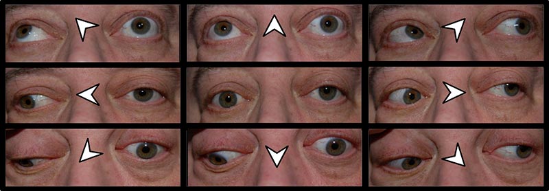
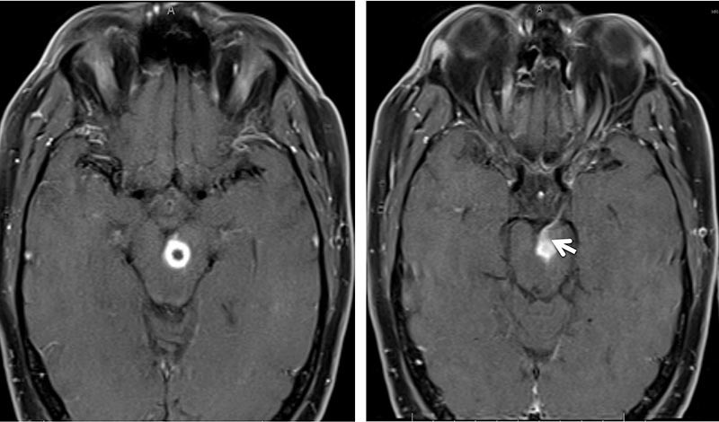
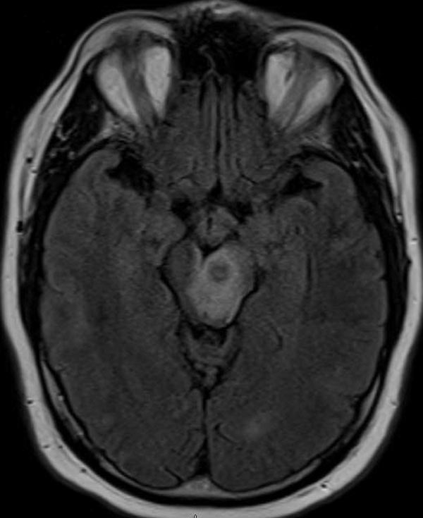
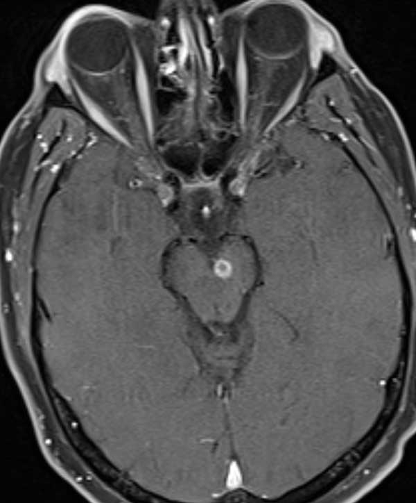
This patient's presentation with a partial progressive pupil-involving third nerve palsy with contralateral hemiplegia was concerning for a midbrain lesion. MRI brain with contrast was performed and revealed a ring-enhancing lesion with associated vasogenic edema in the left side of the midbrain, involving the left third nerve fascicle and medial left cerebral peduncle (Figures 2 and 3). No other intra-axial lesions were identified. MRA head with contrast was unremarkable.

Figure 2: a) Post-gadolinium axial T1-weighted MRI demonstrating a ring-enhancing lesion of the midbrain and b) enhancement of the left third nerve (arrow).
Post-gadolinium axial t1-weighted MRI, see caption for details
Figure 3: Axial FLAIR showing vasogenic edema surrounding the midbrain lesion.
Axial FLAIR, see cpation for details

The patient was admitted to the Neurology service. With guidance from the Infectious Disease team, the patient had a full work-up for infectious, inflammatory, and neoplastic sources which included:

Blood Studies

  • CBC—mild anemia
    • WBC count—5.1 k/mm3; RBC count—3.97 M/mm3 ; hemoglobin—12.7 g/dL; hematocrit—37%; MCV—94 fL; RDW—14.3%
  • WBC differential—left shift
    • Segmented—3320/mm3; lymphocytes—1090/mm3; monocytes—410/mm3; eosinophils—90/mm3; basophils—50/mm3; bands—50/mm3; metamyelocytes—50/mm3; reactive lymphocytes—50/mm3; toxic granulation—present ; hypersegmented neutrophils—present
  • LFT—slightly elevated
    • ALT—113 U/L; AST—50 U/L
  • Basic Metabolic Panel—normal limits
    • Sodium—140 mEq/L; potassium—3.9 mEq/L; chloride—103 mEq/L; CO2—25 mEq/L; anion gap—12 mEq/L; BUN—14 mg/dL ; creatinine—1.3 mg/dL
  • Aerobic/anaerobic cultures—no growth
  • Gram stain—no organisms
  • Aspergillus Galactomannan Antigen Assay—negative
  • Beta-D-Glucan Assay—positive (74 pg/mL)
  • Blastomyces Antibody—negative
  • Coccidioides Antibody—negative
  • EBV PCR—negative
  • ESR—elevated (31 mm/h)
  • CRP—normal limits (<0.5 mg/dL)
  • Histoplasma Antigen Assay—negative
  • IgM/IgA/IgG Serum Assay—normal limits (16 mg/dL; 54 mg/dL; 610 mg/dL, respectively)
  • Kappa-Lambda Free Light Chain Ratio—normal limits (0.29)
  • Quantiferon TB Gold—negative
  • Serum Protein Electrophoresis—normal limits (total protein—6.5 g/dL)

CSF Studies

  • CSF analysis—protein slightly elevated
    • Total protein—49 mg/dL; glucose—65 mg/dL; CSF clarity—clear; total nucleated count—0 cells; RBC count—9 cells; neutrophil count—0 cells
  • Aerobic/anaerobic cultures—no growth
  • Gram stain—no organisms
  • Cytomegalovirus PCR—negative
  • Herpes Simplex PCR—negative
  • Histoplasma Antigen Assay—negative
  • Toxoplasma Antibody—negative
  • Toxoplasma PCR—negative
  • Varicella Zoster PCR—negative

Urine Studies

  • Histoplasma Antigen Assay—negative

Imaging Studies

  • MRI brain with contrast, MRA head with contrast
  • CT chest/abdomen/pelvis with contrast
  • Transesophageal Ultrasound

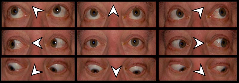
The workup was unrevealing, except for a weakly-positive Beta-D-Glucan Assay, which suggested a fungal etiology. Although a biopsy would have provided significant information regarding the etiology of the lesion, it was considered too risky given the difficult to access location and high likelihood of causing a greater and irreversible neurologic deficit. Therefore, empiric treatment with voriconazole and caspofungin was initiated. The treatment was subsequently changed to posaconazole, as the patient developed severe photosensitivity with the voriconazole (1). Serial Beta-D-Glucan measurements demonstrated a two-fold decease over a two week period. The patient was re-evaluated in the Neuro-Ophthalmology clinic 2 months after the initiation of anti-fungal therapy. During that visit, he showed a dramatic improvement in motility, with full adduction and marginally limited supraduction and infraduction of the left eye (Figure 4). Pupils were equal and briskly reactive to light. Almost complete resolution of his ptosis was also noted.

Figure 4: External photograph demonstrating full abduction and adduction with minimal limitation of supraduction and infraduction of the left eye (2 months after initiating therapy).
External photo, see caption for details
Figure 5: Post-gadolinium axial T1-weighted MRI showing a decrease in the size of the ring-enhancing lesion (5 months after initiating therapy).
MRI, see caption for details

The patient was continued on posaconazole and scheduled for brain MRI with contrast and Neuro-Ophthalmology follow-up in 3 months. MRI demonstrated a drastic reduction in the size of the ring-enhancing lesion and associated vasogenic edema (Figure 5). The radiographic improvement correlated with clinical improvement as well. Motility was noted to be full, except for minimal residual limitation of supraduction. Pupils were equal and briskly reactive to light.

Discussion:

Lesion Localization

Based on his examination findings of a partial, pupil-involving third nerve palsy with contralateral hemiplegia, the lesion could be localized to the midbrain prior to imaging. Once focused to the midbrain, additional signs/symptoms allowed for more specific localization within the midbrain. The presence of a left-sided third nerve palsy and right sided pyramidal signs suggested a left midbrain lesion. Weakness of the right upper extremity with a negative Babinski sign of the right lower extremity indicated involvement of the upper extremity, but sparing the lower extremity. This allowed localization of the lesion to the medial cerebral peduncle, due to its topographical arrangement (upper extremity-medial, lower extremity-lateral). Finally, the posterior boundary of the lesion could be determined by observing whether the medial lemniscus (contralateral loss of two-point discrimination, vibration, and proprioception) or the superior cerebellar peduncle (contralateral ataxia) was involved (2,3). Because the patient did not demonstrate any of these findings, the lesion was localized anterior to the medial lemniscus and the superior cerebellar peduncle. Therefore, the clinical symptoms and signs allowed for localization of the lesion to the left side of the midbrain, involving the medial cerebral peduncle and third nerve fascicle. Note that this patient could also be described as having Weber Syndrome, a third nerve fascicular syndrome occurring due to involvement of the cerebral peduncle and the adjacent third nerve fasciculus (4). Other third nerve fascicular syndromes are listed in the table below.

Ring-Enhancing Lesion Etiology

The differential diagnosis for a ring enhancing lesion in an immunocompromised individual can be separated into three major categories: infectious, neoplastic, or inflammatory (5). During the work-up of this patient, investigations for bacterial, viral, parasitic, inflammatory, and neoplastic etiologies all came back negative. Only the Beta-D-Glucan assay was positive. This assay measures 1,3-Beta-D-Glucan, which is a component of the cell wall of most fungi, excluding Cryptococcus, Zygomycetes, and the yeast form of Blastomyces (sensitivity 50%, specificity 99%) (6,7). Serial measurements of Beta-D-Glucan levels were utilized in order to monitor response to therapy. In this case, a decrease in Beta-D-Glucan levels seemed to correlate with both radiologic and clinical improvement, although there is no current literature addressing the utility of following Beta-D-Glucan levels to monitor patient response to therapy.

Treatment

Because the Beta-D-Glucan Assay suggested that the lesion was a fungal abscess, empiric treatment with voriconazole and caspofungin was initiated. Voriconazole was chosen due to its efficacy against invasive aspergillus and broad-spectrum of anti-fungal coverage. In 2008, the Infectious Disease Society of America advised that voriconazole was superior to amphotericin B for treatment of invasive aspergillus (8). Although there is limited evidence to support the use of voriconazole over amphotericin B for invasive fungal infections other than aspergillus, it was felt to be adequate because of its broad-spectrum of action and less severe side-effects compared with amphotericin B. Combination therapy with caspofungin was also utilized in this case during the first four weeks, even though there are limited studies to suggest that combination therapy is superior to monotherapy (9). During treatment, the patient experienced photosensitivity, a well-characterized side-effect of voriconazole treatment and, thus, was switched to posaconazole. When compared with voriconazole, posaconazole has a similar broad-spectrum of activity, but lacks efficacy against invasive aspergillus. Since CNS fungal infections can be difficult to cure, this patient will remain on posaconazole treatment long-term until radiologic signs of the lesion are no longer evident.

Diagnosis: Third Nerve Palsy with Contralateral Hemiplegia Secondary to Midbrain Fungal Abscess

Epidemiology of a midbrain fungal abscess

Immunocompromised Individuals

  • Use of chronic immunomodulators
  • Stem cell transplant
  • Solid organ transplant
  • Leukemia/Lymphoma
  • Diabetes mellitus
  • HIV/AIDS
  • Malnutrition

Signs of a 3rd nerve palsy from a fascicular midbrain lesion

  • Impaired ocular motility: limited adduction, infraduction, and supraduction (classically “down and out”)
  • Ptosis
  • Impaired levator function
  • Dilated pupil
  • ***3rd nerve palsy can be partial or incomplete
  • Third nerve fascicular syndromes
    • Weber's syndrome: ipsilateral oculomotor nerve palsy with contralateral hemiplegia/hemiparesis due to damage to fascicular oculomotor fibers and motor fibers in the cerebral peduncle.
    • Claude's syndrome: ipsilateral oculomotor nerve palsy with contralateral ataxia due to involvement of the superior cerebellar peduncle.
    • Benedikt's syndrome: ipsilateral oculomotor nerve palsy with contralateral tremor due to involvement of the red nucleus and/or substantia nigra.
    • Nothnagel's syndrome: ipsilateral oculomotor palsy and ipsilateral cerebellar ataxia due to involvement of the brachium conjunctivum.

Symptoms of a 3rd nerve palsy from a fascicular midbrain lesion (10)

  • Diplopia
  • Ptosis
  • Possible weakness, tremor, ataxia

Treatment of a midbrain fungal abscess

Options Include one of the following:

  • Voriconazole (300 mg twice daily) + Caspofungin (50 mg IV x 4 weeks)
  • Posaconazole (200 mg three times daily)

Differential Diagnosis for Ring-Enhancing Lesion in Immunosuppressed:

Infectious

  • Bacterial
    • Pyogenic abscess
    • Tuberculoma
    • Tuberculous Abscess
    • MAI Infection
    • Syphilis
    • Listeriosis
    • Nocardiosis
    • Actinomycosis
  • Fungal
    • Rhodococosis
    • Zygomycosis
    • Histoplasmosis
    • Coccidiomycosis
    • Aspergillosis
    • Mucormycosis
    • Cryptococcosis
  • Parasitic
    • Toxoplasmosis
    • Amebic Brain Abscess
    • Echinococcosis
    • Neurocysticercosis
    • Cerebral sparganosis

Neoplastic

  • Metastases
  • Primary brain tumor
  • Primary CNS lymphoma

Inflammatory

  • Sarcoidosis
  • Multiple Sclerosis
References
  1. McCarthy KL, Playford EG, Looke DF, et al, “Severe Photosensitivity Causing Multifocal Squamous Cell Carcinomas Secondary to Prolonged Voriconazole Therapy,” Clin Infect Dis, 2007;44(5):e55-6.
  2. Bruce BB, Biousse V, Newman NJ: Third nerve palsies. Semin Neurol 2007;27:257.
  3. Duane E. Haines. Neuroanatomy: An Atlas of Structures, Sections, and Systems. 7th. Philadelphia: Lippincott Williams & Wilkins, 2007. p 138.
  4. Liu GT, Crenner CW, Logigian EL, Charness ME, Samuels MA. Midbrain syndromes of Benedikt, Claude, and Nothnagel: setting the record straight. Neurology. 1992;42(9):1820-2.
  5. Ropper AH, Samuels MA. Chapter 32. Infections of the Nervous System (Bacterial, Fungal, Spirochetal, Parasitic) and Sarcoidosis. In: Ropper AH, Samuels MA, eds. Adams and Victor's Principles of Neurology. 9th ed. New York: McGraw-Hill; 2009.
  6. Pickering JW, Sant HW, Bowles CA, Roberts WL, Woods GL. Evaluation of a (1->3)-beta-D-glucan assay for diagnosis of invasive fungal infections. J Clin Microbiol. 2005;43(12):5957.
  7. Karageorgopoulos DE, Vouloumanou EK, Ntziora F, Michalopoulos A, et al. β-D-glucan assay for the diagnosis of invasive fungal infections: a meta-analysis. Clin Infect Dis. 2011;52(6):750.
  8. Infectious Diseases Society of America. Treatment of aspergillosis: clinical practice guidelines of the Infectious Diseases Society of America. Clin Infect Dis. 2008;46(3):327.
  9. Vazquez JA. Clinical practice: combination antifungal therapy for mold infections: much ado about nothing? Clin Infect Dis. 2008;46(12):1889.
  10. Thompson RA: Clinical features of central nervous system fungus infection, in Thompson RA, Green JR (eds): Advances in Neurology. Vol 6. New York, Raven Press, 1974, pp 93-100.
Suggested citation format:

Baldwin A, Thurtell MJ. Third Nerve Palsy with Contralateral Hemiplegia Secondary to Midbrain Fungal Abscess. EyeRounds.org. September 5, 2012. Available from: https://eyerounds.org/cases/156-Fungal-abscess-causing-3rd-nerve-palsy.htm.


last updated: September 5, 2012

Image Permissions:
Creative Commons

Ophthalmic Atlas Images by EyeRounds.org, The University of Iowa are licensed under a Creative Commons Attribution-NonCommercial-NoDerivs 3.0 Unported License.