This site uses tracking information. Visit our privacy policy. Click to agree to this policy and not see this again.

Ophthalmology and Visual Sciences

Pattern Dystrophy Associated with Myotonic Dystrophy

Pattern Dystrophy Associated with Myotonic Dystrophy

Brandon Menke and James C. Folk, M.D.
September 9, 2013

Chief Complaint: “Crinkled” vision in the left eye.

History of Present Illness

A 56-year-old female presented to the University of Iowa Retina Service with a one-year history of increasingly distorted vision in the left eye (OS) described as “crinkled”. She had been previously diagnosed with an epiretinal membrane (ERM) in both eyes (OU) and a lamellar macular hole OS which was observed. Best corrected vision was noted on prior examination to be 20/25 in the right eye (OD) and 20/30 OS. In addition, the patient had a known history of type 1 myotonic dystrophy with mild ptosis OU, progressive lower extremity weakness, shortness of breath and fatigue.

Past Ocular History

  • Pseudophakia OU
  • ERM OU
  • Lamellar macular hole OS
  • Myopia and presbyopia

Past Medical History

  • Myotonic dystrophy (type 1)
  • Asthma
  • Heart arrhythmia
  • Osteoarthritis

Medications

  • Lisinopril
  • Ranitidine

Family History

Non-contributory

Social History

No alcohol or tobacco use

Review of systems

Remarkable for shortness of breath, fatigue, and progressive lower extremity weakness.

Ocular Exam

Visual acuity (without correction):

OD: 20/20-3
OS: 20/60 (pinhole 20/40+2)

Intraocular pressure (IOP):

OU: Normal

Pupils

OU: Round and briskly reactive without afferent pupillary defect

Extraocular motility

OU: Full

Confrontation visual field

OU: Full

External exam

OU: No facial weakness

Slit lamp examination

Lids/Lashes: Mild ptosis OU

Conjunctiva/Sclera: Clear and quiet OU

Cornea: Clear OU

Anterior Chamber: Deep and quiet OU

Iris: Normal architecture OU

Lens: Posterior chamber intraocular lens OU

Fundus examination: Figure 1

Disc: Normal with 0.4 cup to disc ratio OU

Macula:

-OD: 1+ ERM and pigment clumping
-OS: Lamellar macular hole and pigment clumping, faint ERM

Vessels:  Normal OU

Periphery:

-OD: Inferior cobblestone degeneration
-OS: Normal

Ancillary tests: Color fundus photography, Optical coherence tomography (OCT), Fundus autofluorescence (FAF)


Pattern Dystrophy

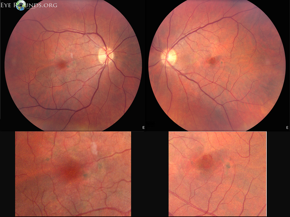
Figure 1. Top: Color fundus photos demonstrate pigment clumping OU, ERM OD, lamellar hole and faint ERM OS. Bottom: Images have been magnified to show macular pigment clumping. The lipofuscin is not well documented on these images.


Optical Coherence Tomography (OCT) images demonstrate an ERM along with hyper-reflective material in the outer retinal layers causing dome-shaped elevations OD

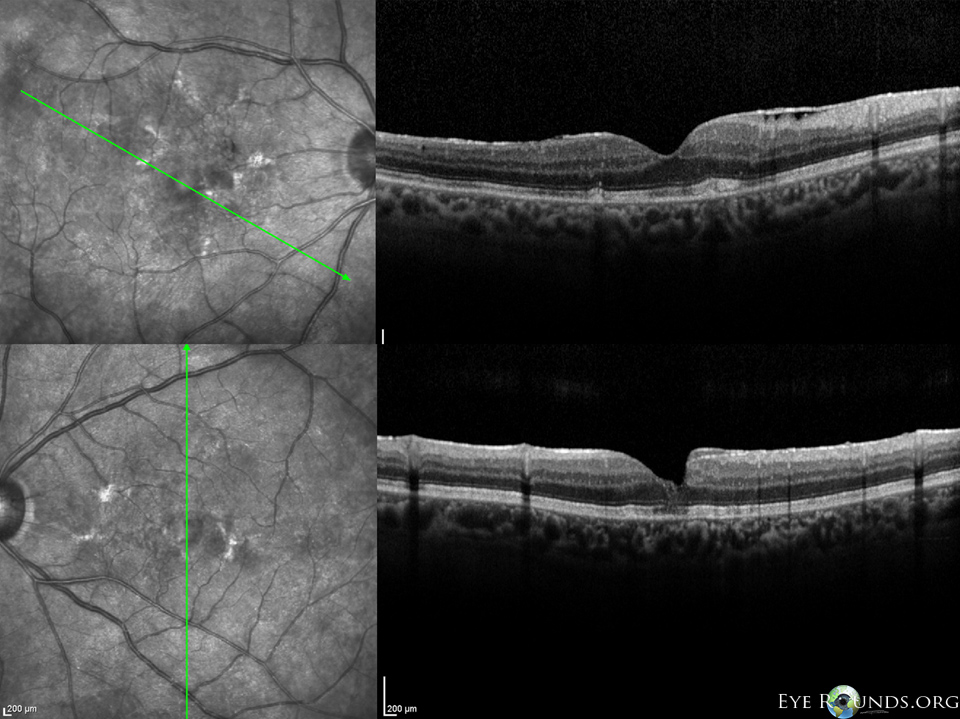
Figure 2. Optical Coherence Tomography (OCT) images demonstrate an ERM along with hyper-reflective material in the outer retinal layers causing dome-shaped elevations OD. These elevations appear to correspond with pigment clumping seen on fundoscopy. In the left eye there is a faint remnant of an ERM, a lamellar macula hole and mild disruption of the photoreceptor layers in the macular center.


Fundus autofluorescence

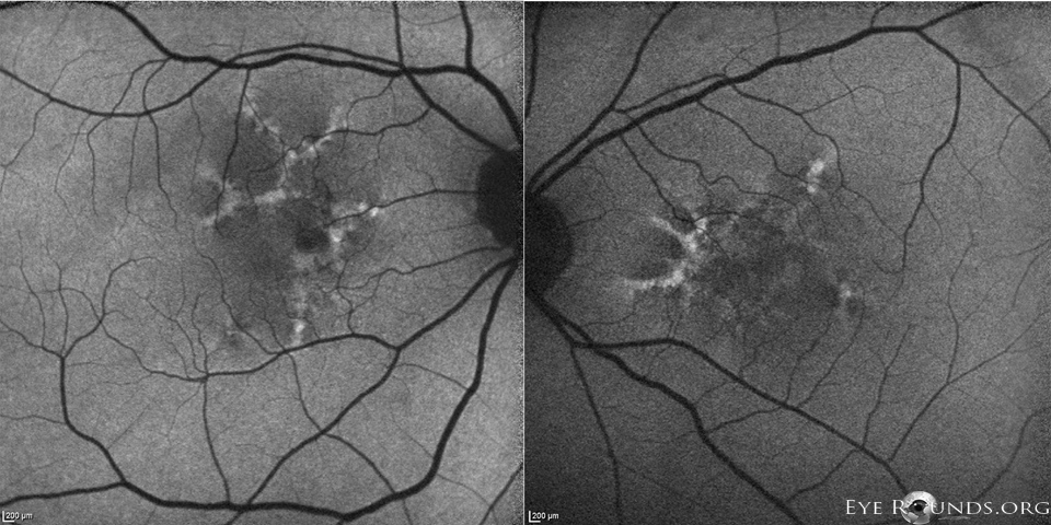
Figure 3. Fundus autofluorescence (FAF) images reveal a hyperfluorescent material, lipofuscin, which is found throughout the macula OU in a butterfly-shaped pattern. Hypofluorescent areas corresponded to pigment clumping seen on fundoscopy.


Final Diagnoses

  • Butterfly-shaped pattern dystrophy
  • Epiretinal membrane OU
  • Lamellar macular hole OS

Summary

The patient is a 56-year-old female with type 1 myotonic dystrophy with known ERM OU and lamellar hole OS who presented with “crinkled” images in her vision OS over the prior year. While her initial symptoms were felt to be mainly due to ERM OU and lamellar hole OS, her further decrease in vision OS was thought to be the result of macular changes associated with pattern dystrophy.

Discussion

Pattern dystrophy (PD) refers to a group of dominantly inherited macular diseases that are characterized by the accumulation of pigment by retinal pigment epithelium (RPE) cells [1,2]. Although currently classified according to the pattern of lipofuscin and pigment deposition seen on funduscopic examination, it is best to view PDs as a single disease with variable penetrance and expressivity. Evidence for lumping the various patterns into a single disease includes families in which members with identical gene mutations express different patterns in the macula, as well as patients that show different patterns in each eye. Furthermore, PDs have been shown to progress into different subtypes of PD [3-6]. The gene implicated to cause pattern dystrophy is the retinal degeneration slow (RDS)/peripherin gene which is located on chromosome 6p21.2. Specific missense mutations include Gly167Asp[7], Arg172Trp[8,9], Cys213Arg[10], Cys213Tyr[11], and Cys250Phe[12], as well as the nonsense mutations found in Gln239ter[13] and Tyr285ter[13]. The RDS gene encodes for a photoreceptor outer-segment disc maintenance glycoprotein that when disrupted, is thought to interfere with the photoreceptor membrane’s integrity [11,14-16]. PD subtypes include butterfly-shaped pattern dystrophy, reticular dystrophy, multifocal PD, simulating flavimaculatus, adult vitelliform dystrophy, and fundus pulverlentus [17]. The patient described in this case displayed the butterfly-shaped pattern on FAF, consistent with butterfly-shaped pattern dystrophy (BPD).

BPD, a well-known subtype of macular pattern dystrophy, displays a unique pattern of hyperfluorescence as well as hypofluorescence on FAF imaging [20]. In a sense, FAF can be thought of as a non-invasive assessment of RPE health where areas of differing fluorescent properties reflect the changes and discrepancies in lipofuscin contained within the RPE. While not shown here, classically BPD will show early hyperfluorescence outlining hypofluroescent regions on fluorescein angiography (FA) [18]. On FAF, lipofuscin will be hyperfluorescent whereas atrophy or pigmentation will be hypoautofluorescent. With this in mind, age-related macular degeneration (AMD) can mimic the appearance of PD preceding the onset of geographic atrophy. Furthermore, the late appearance of PD can appear very similar to AMD as drusen progressively become less prominent. In such cases, distinguishing between AMD and PD should be made with FAF by way of monitoring changes in fluorescence. 

The diagnosis of BPD usually occurs in midlife and patients are typically found to be asymptomatic at the time of diagnosis. Classically, patients with BPD suffer modest losses of vision later in life. However, some patients can develop geographic atrophy and depigmentation in their fifth and sixth decade of life, leading to significantly reduced visual acuity. Furthermore, BPD has been linked to an increased risk for development of choroidal neovascularization, which can also cause further vision loss [19].

Interestingly, PD has been associated with seemingly unrelated diseases, namely myotonic dystrophy [21]. Myotonic dystrophy is a highly variable, multisystem, slowly progressing disease that is classically characterized by the wasting of muscles. Type 1 myotonic dystrophy, also known as Steinert disease, is an autosomal dominant disease caused by a trinucleotide repeat of CTG sequences in the dystrophia myotonica protein kinase (DMPK) gene, located on chromosome 19. Immediately downstream of DMPK is SIX5, a gene thought to play a part in eye development. The CTG repeat sequences are located within the promoter of SIX5 and may presumptively be a component for the etiology of the ocular manifestations associated with myotonic dystrophy [22]. While the exact genetic etiology for the link between myotonic dystrophy and pattern dystrophy remains unclear, this patient’s case further demonstrates their association.

Differential Diagnosis

  • Best disease
  • Age-related macular degeneration
  • Central serous retinopathy
  • Pigment epithelial detachment
  • Stargardt disease
  • Dominant drusen (Malattia Leventinese)

Epidemiology

  • The incidence is unknown as the majority of patients are asymptomatic and it can be difficult to differentiate from age-related macular degeneration. It appears that PD is an uncommon but not rare condition
  • Associated with myotonic dystrophy and pseudoxanthoma elasticum. The latter association, however, may be due to a pattern-like appearance in fundi with angioid streaks
  • Because of the variable penetrance and expressivity, family members with identical mutations have been observed to have different patterns of pigment accumulation. Furthermore, some individual patients have developed different PDs over time, as well as having different PDs in each eye


Symptoms

  • The age of onset is highly variable for pattern dystrophy
    • Many patients remain asymptomatic
    • The majority of patients who do suffer from decreased vision often experience symptoms in the fifth or sixth decade of life. Most patients will only suffer from mild impairment of their central vision


Signs

  • Lipofuscin deposits in various patterns along with pigmentation in macula
  • Minority of patients will present with central visual loss or disturbances and possible visual distortions
  • Rarely, the disease can progress to RPE atrophy or to choroidal neovascularization
  • PD patients have normal dark adaptation, color vision, and intact peripheral fields
  • Pattern dystrophy may be misdiagnosed as Age-related macular degeneration (AMD) because well-developed PD will display drusen-like deposits of lipofuscin accumulation


Treatment

  • There is currently no treatment available for this macular disease
  • Visual aids should be considered for individuals with vision loss
  • Use of Amsler grid and other central vision monitoring to detect CNV
  • Anti-vascular endothelial growth factor agents for associated CNV


References
  1. Marmor MF, Byers B. Pattern Dystrophy Of The Retinal Pigment Epithelium. Am J Ophthalmol. 1976;13:112-116.
  2. Hsieh RC, Fine BS, Lyons JS. Pattern Dystrophy Of The Retinal Pigment Epithelium. Arch Ophthalmol. 1977;95:429-435.
  3. Kim RY, Dollfus H, Keen TJ, et al. Autosomal Dominant Pattern Dystrophy of the Retina Associated with a 4-basepair insertion at Codon 140 in the Peripherin/RDS gene. Arch Ophthalmol. 113, 451-455.
  4. Yang Z, Li Y, Jiang L, Karan G, et al. A novel RDS/Peripherin gene mutation Associated with Diverse Macular Phenotypes. Ophthalmic Genet. 25:133-145.
  5. Fossarello M, Bertini C, Galantuomo MS, et al. Deletion in the Peripherin/RDS gene in Two Unrelated Sardinian Families with Autosomal Dominant Butterfly-Shaped Macular Dystrophy. Arch Ophthalmol. 1996;114:448-456.
  6. Watzke RC, Folk JC, Lang RM. Pattern Dystrophy Of The Retinal Pigment Epithelium. Ophthalmology. 1982;89:1400-1406.
  7. Nicholas BE, Sheffield VC, Vanderburgh K, et al. Butterfly-Shaped Pigment Dystrophy of the Fovea Caused by a point Mutation in codon 167 of the RDS gene. Nat Genet. 1993;3:202-207.
  8. Downes SM, Fitzke FW, Holder GE, et al. Clinical Features of Codon 172 RDS Macular Dystrophy: Similar Phenotype in 12 Families. Arch Ophthalmol. 1999;117:1373-1383.
  9. Wells J, Wroblewski J, Keen J, et al. Mutations in the Human Retinal Degeneration Slow (RDS) gene Can Cause Either Retinitis Pigmentosa or Macular Dystrophy. Nat Genet. 1993;3:213-218.
  10. Payne AM, Downes SM, Bessant DA, et al. Founder Effect Seen in the British Population of the 172 Peripherin/RDS Mutation and Further Refinement of the Genetic Position of the Peripherin/RDS. Am J Hum Genet. 1998;62:192-195.
  11. Zhang K, Garibaldi DC, Li Y, et al. Butterfly-Shaped Pattern Dystrophy: A Genetic, Clinical, and Histopathololgical report. Ophthlmic Mol Genet. 2002;120:485-490.
  12. Francis PJ, Schultz DW, Gregory AM, et al. Genetic and Phenotypic Hetrogeneity in Pattern Dystrophy. Br J Ophthalmol. 2005;89:1115-1119.
  13. Kohl S, Christ-Adler M, Apfelstedt-Sylla E, et al. RDS/Peripherin Gene Mutations are Frequent Causes of Central Retinal Dystrophies. J Med Genet. 1997;34:620-626.
  14. Arikawa K, Molday LL, Molday RS, Williams DS. Localization of RDS/Peripherin in the Disk Membranes of Cone and Rod Photoreceptors: Relationship to Disk Membrane Morphogenesis and Retinal Degeneration. J Cell Biol. 1992;116:659-667.
  15. Connell G, Bascom R, Molday L, et al. Photoreceptor Peripherin is the Normal Product of the gene Responsible for Retinal Degeneration in the RDS Mouse. Proc Natl Acad Sci U S A. 1991;88:723-726.
  16. Travis GH, Sutcliffe JG, Bok D. The Retinal Degeneration Slow (RDS) Gene Product is a Photoreceptor Disc Membrane-associated Glycoprotein. Neuron. 1991;6:61-70.
  17. Ayazi S, Fagan R. Pattern Dystrophy of Pigment Epithelium. Retina. 1981;1:287-289.
  18. Tuppuranen K, Mantyjarvi M. The importance of fluorescein angiography in diagnosing pattern dystrophies of the retinal pigment epithelium. Doc Ophthalmol. 1994;87:233-243.
  19. Theodoros Empeslidis, Athanasios Vardarinos, James Deane, and Somnath Banerjee. (2012). Intravitreal Ranibizumab in the Treatment of Butterfly-Shaped Pattern Dystrophy Associated with Choroidal Neovascularization: A Case Report. Case Reports in Ophthalmology.
  20. Holz FG, Bellman C, Staudt, S, et al. Fundus autofluorescence and development of geographic atrophy in age-related macular degeneration. Invest Ophthalmol Vis Sci 2001;42:1051-1056.
  21. Louprasong AC, Light DJ, Diller RS. Spider Dystrophy as an ocular manifestation of myotonic dystrophy. Optometry. 2010 Apr;81(4):188-93.
  22. Winchester CL, Ferrier RK, Sermoni A, Clark BJ, Johnson KJ. Characterization of the expression of DMPK and SIX5 in the human eye and implications for pathogenesis in myotonic dystrophy. Hum Mol Genet. 1999 Mar;8(3):481-92.
Suggested citation format:

Menke B, Folk JC. Pattern Dystrophy Associated with Myotonic Dystrophy. EyeRounds.org. September 9, 2013; available from https://eyerounds.org/cases/179-pattern-dystrophy.htm


last updated: 9/9/2013

Image Permissions:
Creative Commons

Ophthalmic Atlas Images by EyeRounds.org, The University of Iowa are licensed under a Creative Commons Attribution-NonCommercial-NoDerivs 3.0 Unported License.