Chief Complaint: Growth on eyelid
A 94-year-old female is referred for an evaluation for possible skin cancer on the nasal aspect of her left lower lid. The lesion has been present on the left lower lid for about one month. It has grown rapidly in size over the previous two weeks and is now obstructing her vision. She denies any pain, flaking or itching, but the mass does bleed when manipulated.
Multiple skin cancers removed from her face and hands.

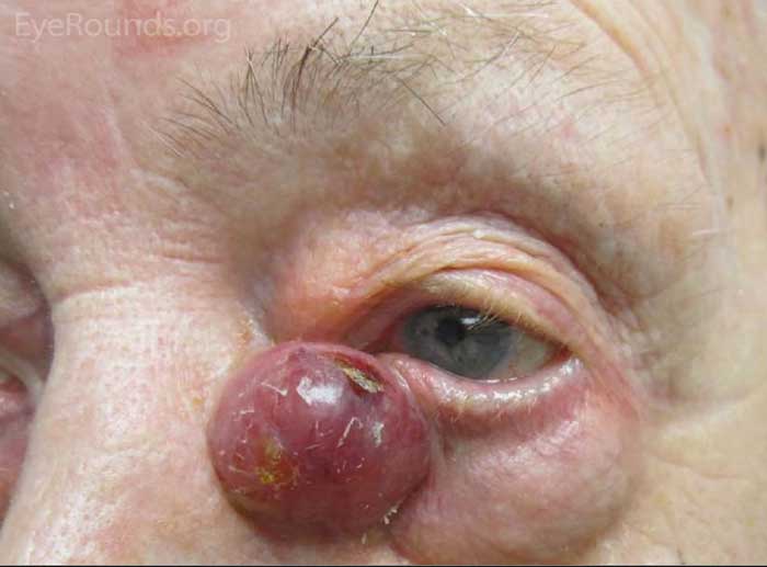
Figure 1: A large lesion arising from the left medial canthus, flesh-colored with a violaceous hue, nearly all inferior to the medial canthal tendon.
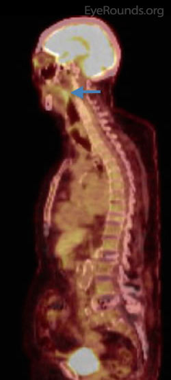
The patient underwent biopsy of the lesion by the dermatology team, which returned with a diagnosis of Merkel cell carcinoma. A subsequent positron emission tomography (PET) scan revealed an enhancing lesion near the inferior border of the ipsilateral parotid gland (Figure 2). Excision was performed with frozen section controls in the operating room followed by a sentinel lymph node dissection including removal of lymph nodes in the parajugular area with the otolaryngology team. The patient then underwent reconstruction of the left medial canthus and medial eyelid defect with a median forehead flap, periosteal strip, and Mustarde advancement flap. Her postoperative course was complicated by postoperative hematoma, which resolved with conservative management. A few months later, the patient underwent focal radiation therapy. She did well following her treatment with no signs of recurrence.

Figure 2: Whole body PET/CT scan showing parotid gland uptake consistent with metastases of the tumor
Merkel cell carcinoma (MCC) is a rare neuroendocrine tumor. These tumors are quite aggressive with high metastatic and mortality rates. The immunohistochemistry and structural features are unique. Recent literature contends that these tumors often exhibit non-epithelial cell and sarcomatous features, which suggests a stem cell origin (1). There are approximately 800-1500 new cases of MCC in the US per year (2). The incidence of MCC has increased from 0.15 cases per 100,000 in 1986 to 0.44 cases per 100,000 in 2001. During this period, the incidence of MCC increased 8.08% per year. In comparison, melanoma increased at a rate of 3.03% each year over a similar time period (3). Furthermore, MCC carries an overall mortality rate of 33% with 5-year survival estimated at 30-64%, compared to an overall mortality of 15% for cutaneous melanoma (4).
In a population based study examining 3870 cases of MCC, the majority of cases were found in Caucasian patients at 94.9% with only 1% of cases diagnosed in African-Americans. Additionally, 61.5% of cases were found in men and 38.5% were found in women (1). A cohort study of 195 patients diagnosed with MCC in the US found a median age at diagnosis of 69, ranging from 34 to 97 years old. Interestingly, extensive immunosuppression was found in 7.8% of this cohort, including chronic lymphocytic leukemia, HIV, and organ transplantation. At initial presentation, 34.7% presented with nodal disease and 5.9% presented with distant metastatic disease (5).
Clinically, MCC can be confused with a variety of dermatologic lesions including cystic/acneiform lesions, lipoma, non-melanoma skin carcinoma, and lymphoma. Furthermore, 97.6% of MCC present as cutaneous lesions, most commonly on the head and neck (26.9%) (1). The five most common features of MCC at presentation are asymptomatic or lack of tenderness (88%), rapid expansion (63%), immunosuppression (7.8%), older than 50 years of age (90%), and location on UV exposed skin (81%); this lead to the development of the acronym AEIOU to aid in diagnosis. In the aforementioned cohort of 195 patient with MCC, 89% met ≥ 3 features, 32% met ≥ 4 features, and 7% met all 5 features (5). However, biopsy and immunohistochemistry are required to establish the diagnosis (3). PET/CT is frequently used to evaluate the extent of disease and to help guide treatment.

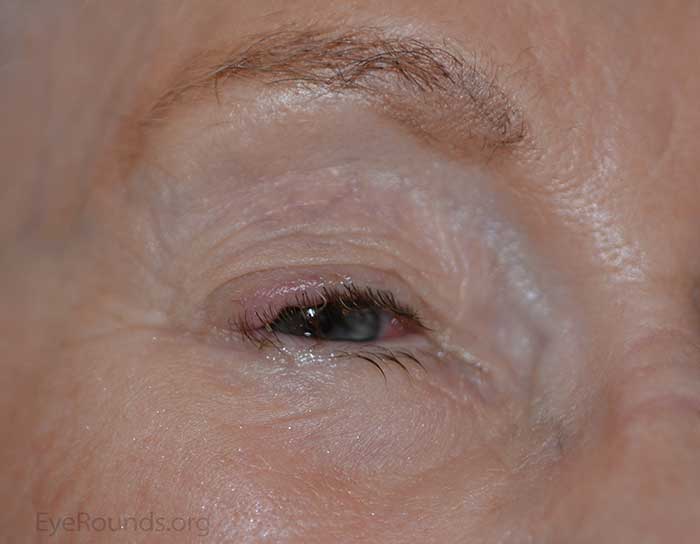
Figure 3: Another example of Merkel cell carcinoma presenting as a flesh-colored papule arising from the superotemporal portion of the upper eyelid.
Prognosis is heavily influenced by the stage and location of disease. The Memorial Sloan-Kettering Cancer Center (MSKCC) introduced the most widely used staging system in 2005 and was adopted by the National Cancer Institute (3). The 10-year survival rate was greater in women than men at 64.8% and 50.5%, respectively. Patients greater than 70 years of age had the lowest 10-year survival rate at 52.7%. Those less than 49 years of age had the highest 10-year survival rate at 59.1%. Disease located on the upper extremities had the best prognosis with 60.7% survival at 10 years. Furthermore, tumors less than 2 cm had a 10-year survival rate of 61.0%. Conversely, tumors greater than 2 cm had a 10-year survival rate of 39.6%. It is also important to note that immunosuppression correlates with poorer outcomes (5, 6, 7).
Localized disease can be treated with surgical excision and adjuvant radiotherapy (8, 9). During excision, wide margins are recommended with a 1 cm margin for lesions less than 2 cm in size and a 2 cm margin for lesions larger than 2 cm (9). Mohs can be a considered for smaller lesions (9). Surgical excision with adjuvant radiotherapy was 3.7 times less likely to develop recurrent local disease and 2.9 times less likely to develop regional disease. Treatment of nodal disease has transitioned in recent years from complete lymph node dissection to sentinel lymph node biopsy followed by surgical excision with adjuvant radiotherapy. It has been recommended that all Merkel cell carcinomas should undergo sentinel lymph node biopsy at the time of excision. Metastatic disease requires further evaluation with personalized management consisting of palliative surgeries, radiotherapy, and chemotherapy.
Merkel cell carcinoma is a rare, but aggressive disease that has been increasing in incidence. A high suspicion of MCC is necessary for prompt diagnosis and can be strengthened with use of the AEIOU mnemonic. Prognosis is generally poor with over two times the mortality rate of melanoma. Treatment is based on staging of disease. Localized disease is treated with localized excision with wide margins and adjuvant radiotherapy. Nodal disease requires further evaluation via sentinel lymph node biopsy in addition to surgical excision and radiotherapy, whereas metastatic disease requires more extensive management.
EPIDEMIOLOGY
|
SIGNS
|
SYMPTOMS
|
TREATMENT
|
Lenci L, Chung A, Allen RC. Merkel Cell Carcinoma of the Eyelid. EyeRounds.org. posted August 24, 2015; Available from: http://www.EyeRounds.org/cases/217-Merkel-Cell-Carcinoma-Eyelid.htm

Ophthalmic Atlas Images by EyeRounds.org, The University of Iowa are licensed under a Creative Commons Attribution-NonCommercial-NoDerivs 3.0 Unported License.