This site uses tracking information. Visit our privacy policy. Click to agree to this policy and not see this again.

Ophthalmology and Visual Sciences

Laser Pointer Maculopathy

Laser Pointer Maculopathy

Mahsaw Motlagh, MD; Mark Wilkinson, OD, FAAO
March 25, 2021

INITIAL PRESENTATION

Chief Complaint: Black spot in the right eye

History of Present Illness:

The patient was a 17-year-old previously healthy female who presented with acute vision loss in the right eye after playing with a sibling at home. Her vision loss occurred suddenly following a handheld laser pointer being directed into both eyes. She presented to clinic one week after her initial injury as her vision loss had not resolved. On evaluation she noted a persistent "big black spot" in the center of her vision in the right eye, with an associated headache when focusing on fine detail. She denied photophobia, metamorphopsia, pain with eye movements, flashing lights,

Past Ocular History:

  • None

Medical History:

  • Attention deficit hyperactivity disorder (ADHD)
  • Generalized anxiety disorder
  • Major depressive disorder

Medications:

  • Aripiprazole
  • Buspirone
  • Oxcarbazepine
  • Sertraline

Family History:

  • Non-contributory

Social History:

  • Non-contributory

Review of Systems:

  • Negative except as detailed in the history of present illness


OCULAR EXAMINATION

  • Uncorrected Visual Acuity:
    • Right eye (OD): 20/100, no improvement with refraction
    • Left eye (OS): 20/20
  • Ocular Motility/Alignment:
    • Normal in both eyes (OU)
  • Pupils:
    • OD: 4 mm in dark, 2 mm in light, no relative afferent pupillary defect (RAPD)
    • OS: 4 mm in dark, 2 mm in light, no RAPD
  • Confrontational visual fields:
    • OD: Full peripheral fields
    • OS: Full peripheral fields
  • Slit lamp exam:
    • Lids/Lashes: Normal OU
    • Conjunctiva/Sclera: White and quiet OU
    • Cornea: Clear and quiet OU
    • Anterior chamber: Deep and quiet OU
    • Iris: Normal architecture OU
    • Lens: Clear OU
  • Dilated fundus examination (DFE):
    • Vitreous: Clear OU
    • Disc: Normal OU
    • Cup-to-disc ratio: 0.2 OU
    • Macula:
      • OD: Fine line of yellow-white scarring superotemporally that involves the foveal center with associated hyperpigmentation
      • OS: Two fine scars abutting but not directly involving the foveal center
    • Vessels: Normal course and caliber OU
    • Periphery: Normal OU
  • Color fundus photograph of the right macula.
    Figure 1: Color fundus photograph of the right macula (A) demonstrates well-demarcated, punctate chorioretinal scars intersecting the fovea. Color fundus photograph of the left macula (B) demonstrates similar chorioretinal scars superior and nasal to fovea.
     OCT of the right macula showing disruption of the ellipsoid zone with RPE loss involving the umbo with a collection of focal hyperreflective material.
    Figure 2: OCT of the right macula showing disruption of the ellipsoid zone with RPE loss involving the umbo with a collection of focal hyperreflective material.
     OCT of the left macula showing parafoveal ellipsoid zone disruption.
    Figure 3: OCT of the left macula showing parafoveal ellipsoid zone disruption.

    Differential Diagnosis:


    CLINICAL COURSE

    After being evaluated by an outside provider, the patient was referred to the vision rehabilitation service at the University of Iowa. Subjective history corroborated by the patient and family indicated that the patient was playing with a high-powered laser at the time the injury occurred. She was unsure of the power of the laser but acknowledged that her sibling pointed the laser directly into her eyes. Based on the clinical findings and additional testing, a diagnosis of laser pointer maculopathy was made. The patient was counseled extensively on the diagnosis and prognosis of this condition and was given return precautions should she develop progressive vision changes or metamorphopsia.


    DIAGNOSIS: Laser pointer maculopathy


    DISCUSSION

    Etiology/Epidemiology

    Laser pointer maculopathy is the result of laser injury to the retina. Though most frequently reported as a cause of unilateral vision loss, bilateral vision loss can occur with bilateral exposure [1-3]. Unfortunately, with the increasing availability of laser pointers, there has been a corresponding rise in reported ocular laser injury. Since 2014, worldwide cumulative reported laser injuries has more than doubled [3]. Though the recovery of laser pointer maculopathy is generally favorable, recovery to 20/20 vision has declined from 2010 to 2017. This may be due in part to the accessibility of higher laser powers to the general public [3]. Young men are most frequently affected by handheld laser pointer injuries [5], as well as those with learning or mental health issues [6].

    In the United States, laser products are classified into six categories by the Occupational Safety and Health Administration (OSHA): Class I, Class IA, Class II, Class IIIA, Class IIIB, and Class IV [7]. These categories as well as their power and ocular hazard risk are summarized in Table 1. Despite the growing public health risk [8], only twelve states currently have state-level laser regulations in addition to the federal provisions provided under the Radiation Control for Health and Safety Act of 1968 [7]. As a result, there are several reported cases of misclassified lasers sold with incorrect labeling that lead to inadvertent retinal damage [6].

    Table 1. Laser Pointer Classification in the United States and Summary of Hazards [5,7]. (μW = microwatt, mW = milliWatt, sec = seconds, OSHA = Occupational Safety and Health administration)

    Class

    Power

    Direct Ocular Hazard

    OSHA Labeled Hazard

    I

    0.4 μW

    None

    None

    IA

    40 μW

    After >1000 sec of exposure

    "Do not directly view with optical instruments"

    II

    <1 mW

    After >0.25 sec of exposure

    "Do not stare into beam"

    IIIA

    1-5 mW

    With any exposure

    CAUTION "Avoid direct eye exposure"

    IIIB

    5-500 mW

    With any exposure

    WARNING "Avoid exposure to beam"

    IV

    >500 mW

    With any exposure

    DANGER "Avoid eye or skin exposure to direct or scattered radiation"

    Signs/Symptoms

    Patients may be asymptomatic initially but invariably present with blurry vision, central or paracentral scotomata, or metamorphopsias. The base eye exam should be conducted in a routine manner as anterior segment pathology, namely corneal punctate epitheliopathy, is also a well-known sequela of ocular laser injury [13]. In one case of repeated laser injury, superficial iris atrophy was also reported [14].

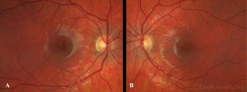
    Fundoscopic examination shows yellow-white lesions or streaks, which may involve the foveal center. There may also be areas of hyper- or hypopigmentation, RPE mottling, and an attenuated foveal reflex. In some cases, patients with self-inflicted laser injuries seem to have scarring with a streak-like appearance, while injuries caused by others tend towards more focal scarring with closer proximity to the fovea [15]. Other posterior findings may include retinal hemorrhage, macular edema, choroidal neovascularization, epiretinal membrane (ERM), or a full-thickness macular hole [16-19]. Following resolution of the initial injury, the macula has a variable appearance ranging from normal to permanent scarring.

    Testing/Imaging

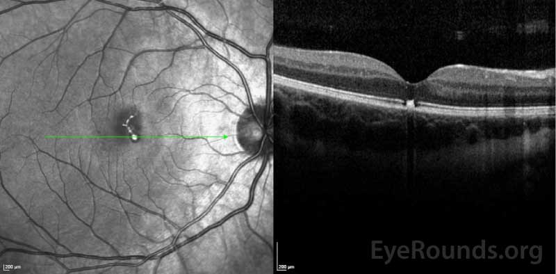
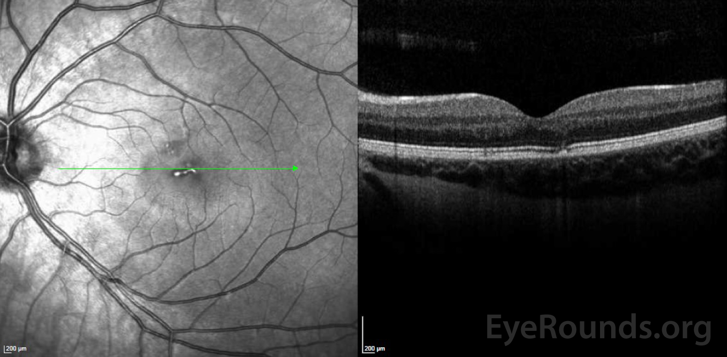
    Amsler grid testing during the initial encounter is a quick, non-invasive method of evaluating a central scotoma. In patients with suspected laser maculopathy, optical coherence tomography (OCT) should be performed. There is often profound focal disruption of the ellipsoid zone and RPE loss on OCT which, in conjunction with a classic history, are diagnostic of laser maculopathy [18, 20, 21]. Vertical hyperreflective bands involving the outer retinal layers may also occur [22-24]. Fluorescein angiography, if available, demonstrates delayed foveal hyperfluorescence and RPE window defects [25-27]. Fundus photography, while not necessary for diagnosis, demonstrates irregular pigmentary changes and RPE disruption as seen in our patient.

    In the absence of a classic history, full-field and multifocal electroretinography (ERG) can objectively quantify retinal function. Preservation of retinal function beyond the focal injury is consistent with laser maculopathy [28]. There is also reported utility of adaptive optics scanning laser ophthalmoscopy (AOSLO) to further characterize photoreceptor damage, though this is seldom available in clinical practice [29].

    Treatment/Management/Guidelines

    Management of laser pointer maculopathy is expectant. Fortunately, the prognosis is favorable, and most patients recover over several months [2, 11, 30]. Those with significant injury, however, can have long-term visual deficits or scotoma. Lasers with a power >5 milliwatts carry a far worse long-term prognosis [3]. There are several studies that advocate oral corticosteroid use as this is thought to reduce the cellular inflammatory response induced by the laser [1 ,5 ,24 ,31 ,32]; however, larger prospective studies are required to demonstrate its clinical utility and effectiveness.

    Patients who develop a macular hole or ERM should be managed according to the degree of visual impairment, with consideration given to the extent of injury and visual prognosis. Choroidal neovascular membranes (CNVM) may be managed with anti-vascular endothelial growth factor agents. If the extent of damage is visually debilitating, referral to a vision rehabilitation specialist may be considered. In all cases, preventative counseling should be undertaken to prevent repeated injury.

    EPIDEMIOLOGY OR ETIOLOGY

    • Cumulative incidence has more than doubled since 2014
    • Young age, male sex, and behavioral disturbances may be risk factors
    • Can be caused by Class IA or higher lasers

      EPIDEMIOLOGY

    • Mechanical stretching of the eye leads to stretching and thinning of the sclera, choroid, and retina that leave them prone to deformation

    SIGNS

    • Discrete macular scarring, unilateral or bilateral
    • Ellipsoid zone disruption and RPE destruction on OCT
    • Window defects on FA

    SYMPTOMS

    • Wide range
    • Asymptomatic
    • Painless central vision loss
    • Vision blurring
    • Metamorphopsia

    TREATMENT/MANAGEMENT

    • No proven effective therapy
    • Prognosis is generally good unless there is significant foveal involvement
    • Management of sequelae (ERM, macular hole, CNVM) as indicated

    References

    • 1. Raevis J, Shrier E. Pediatric Bilateral Blue Laser Pointer-Induced Maculopathy. Case Reports in Ophthalmology 2017;10.1159/000460289. DOI: 10.1159/000460289
    • 2. Chen X, Dajani OAW, Alibhai AY, Duker JS, Baumal CR. LONG-TERM VISUAL RECOVERY IN BILATERAL HANDHELD LASER POINTER–INDUCED MACULOPATHY. RETINAL Cases & Brief Reports 2019;10.1097/icb.0000000000000845. DOI: 10.1097/icb.0000000000000845
    • 3. Torp-Pedersen T, Welinder L, Justesen B, et al. Laser pointer maculopathy – on the rise? Acta Ophthalmologica 2018;10.1111/aos.13856.  https://PubMed.gov/30280504 . DOI: 10.1111/aos.13856
    • 4. Yiu G, Itty S, Toth CA. Ocular safety of recreational lasers. JAMA Ophthalmology, 2014.
    • 5. Lee GD, Baumal CR, Lally D, Pitcher JD, Vander J, Duker JS. Retinal injury after inadvertent handheld laser exposure. Retina 2014;10.1097/IAE.0000000000000397.  https://PubMed.gov/25380069 . DOI: 10.1097/IAE.0000000000000397
    • 6. Linton E, Walkden A, Steeples LR, et al. Retinal burns from laser pointers: a risk in children with behavioural problems. Eye (Basingstoke) 2019;10.1038/s41433-018-0276-z.  https://PubMed.gov/30546136 . DOI: 10.1038/s41433-018-0276-z
    • 7. Administration USOSaH. OSHA technical manual.
    • 8. Alda J, Gómez Sanz F, González Martín-Moro J. Laser pointer maculopathy. A new public health problem? Archivos de la Sociedad Española de Oftalmología (English Edition) 2017;10.1016/j.oftale.2016.11.006. DOI: 10.1016/j.oftale.2016.11.006
    • 9. King A, Gottlieb E, Brooks DG, Murphy MP, Dunaief JL. Mitochondria-derived Reactive Oxygen Species Mediate Blue Light–induced Death of Retinal Pigment Epithelial Cells¶. Photochemistry and Photobiology 2004;10.1562/le-03-17.1.  https://PubMed.gov/15191057 . DOI: 10.1562/le-03-17.1
    • 10. Youssef PN, Sheibani N, Albert DM. Retinal light toxicity. Eye, 2011
    • 11. Barkana Y, Belkin M. Laser eye injuries. Survey of Ophthalmology 2000;10.1016/S0039-6257(00)00112-0.  https://PubMed.gov/10906379 . DOI: 10.1016/S0039-6257(00)00112-0
    • 12. Robertson DM, McLaren JW, Salomao DR, Link TP. Retinopathy from a green laser pointer: A clinicopathologic study. Archives of Ophthalmology 2005;10.1001/archopht.123.5.629.  https://PubMed.gov/15883281 . DOI: 10.1001/archopht.123.5.629
    • 13. Sethi CS, Grey RHB, Dean Hart C. Laser pointers revisited: A survey of 14 patients attending casualty at the Bristol Eye Hospital. British Journal of Ophthalmology 1999;10.1136/bjo.83.10.1164. DOI: 10.1136/bjo.83.10.1164
    • 14. Dolz-Marco R, Souza EC, Iida T, et al. Iris atrophy: A novel sign of repeated selfinflicted laser pointer maculopathy. Retina, 2017
    • 15. Bhavsar KV, Wilson D, Margolis R, et al. Multimodal imaging in handheld laser-induced maculopathy. American Journal of Ophthalmology 2015;10.1016/j.ajo.2014.10.020.  https://PubMed.gov/25448992 . DOI: 10.1016/j.ajo.2014.10.020
    • 16. Fujinami K, Yokoi T, Hiraoka M, Nishina S, Azuma N. Choroidal neovascularization in a child following laser pointer-induced macular injury. Japanese Journal of Ophthalmology, 2010
    • 17. Sun Z, Wen F, Li X, Wu D. Early subfoveal choroidal neovascularization secondary to an accidental stage laser injury. Graefe's Archive for Clinical and Experimental Ophthalmology 2006;10.1007/s00417-005-0169-2.  https://PubMed.gov/16331483 . DOI: 10.1007/s00417-005-0169-2
    • 18. Mtanes K, Mimouni M, Zayit-Soudry S. Laser pointer-induced maculopathy: More than meets the eye. Journal of Pediatric Ophthalmology and Strabismus 2018;10.3928/01913913-20180405-01.  https://PubMed.gov/29913024 . DOI: 10.3928/01913913-20180405-01
    • 19. Alsulaiman SM, Alrushood AA, Almasaud J, et al. High-power handheld blue laser-induced maculopathy: The results of the king khaled eye specialist hospital collaborative retina study group. Ophthalmology 2014;10.1016/j.ophtha.2013.09.006.  https://PubMed.gov/24184164 . DOI: 10.1016/j.ophtha.2013.09.006
    • 20. Rusu I, Sherman J, Gallego-Pinazo R, Lam M, Freund KB. Spectral-domain optical coherence tomography and fundus autofluorescence findings in a case of laser pointer-induced maculopathy. Retinal Cases and Brief Reports 2013;10.1097/ICB.0b013e3182965291. DOI: 10.1097/ICB.0b013e3182965291
    • 21. Zhao N, Liu L. Long-term changes in optic coherence tomography in a child with laser pointer maculopathy: A case report and mini review. Photodiagnosis and Photodynamic Therapy 2017;10.1016/j.pdpdt.2017.03.012.  https://PubMed.gov/28347866 . DOI: 10.1016/j.pdpdt.2017.03.012
    • 22. Tomasso L, Benatti L, La Spina C, et al. Optical coherence tomography angiography findings in laser maculopathy. European Journal of Ophthalmology 2017;10.5301/ejo.5000844.  https://PubMed.gov/28009410 . DOI: 10.5301/ejo.5000844
    • 23. Sánchez-Barahona C, González-Martín-Moro J, Zarallo-Gallardo J, Lozano Escobar I, Cobo-Soriano R. Early changes in optic coherence tomography in a child with laser pointer maculopathy. Archivos de la Sociedad Española de Oftalmología (English Edition) 2017;10.1016/j.oftale.2016.09.013. DOI: 10.1016/j.oftale.2016.09.013.
    • 24. Hossein M, Bonyadi J, Soheilian R, Soheilian M, Peyman GA. SD-OCT features of laser pointer maculopathy before and after systemic corticosteroid therapy. Ophthalmic surgery, lasers & imaging : the official journal of the International Society for Imaging in the Eye 2011;10.3928/15428877-20111208-03.  https://PubMed.gov/22165950 . DOI: 10.3928/15428877-20111208-03
    • 25. Zamir E, Kaiserman I, Chowers I. Laser pointer maculopathy. American Journal of Ophthalmology 1999;10.1016/S0002-9394(99)00017-3.  https://PubMed.gov/10372889 . DOI: 10.1016/S0002-9394(99)00017-3
    • 26. Sell CH, Bryan JS. Maculopathy from handheld diode laser pointer. Archives of Ophthalmology 1999;10.1001/archopht.117.11.1557.  https://PubMed.gov/10565530 . DOI: 10.1001/archopht.117.11.1557
    • 27. Combillet F, Saunier V, Rougier MB, Delyfer MN, Korobelnik JF. Multimodal imaging in a case of self-inflicted laser-induced maculopathy. European Journal of Ophthalmology 2016;10.5301/ejo.5000825.  https://PubMed.gov/27445068 . DOI: 10.5301/ejo.5000825
    • 28. Hanson JVM, Sromicki J, Mangold M, Golling M, Gerth-Kahlert C. Maculopathy following exposure to visible and infrared radiation from a laser pointer: a clinical case study. Documenta Ophthalmologica 2016;10.1007/s10633-016-9530-5.  https://PubMed.gov/26921203 . DOI: 10.1007/s10633-016-9530-5
    • 29. Sheyman AT, Nesper PL, Fawzi AA, Jampol LM. Adaptive optics imaging in laser pointer maculopathy. Ophthalmic Surgery Lasers and Imaging Retina 2016;10.3928/23258160-20160808-14.  https://PubMed.gov/27548458 . DOI: 10.3928/23258160-20160808-14
    • 30. Weng CY, Baumal CR, Albini TA, Berrocal AM. Self-induced laser maculopathy in an adolescent boy utilizing a mirror. Ophthalmic Surgery Lasers and Imaging Retina 2015;10.3928/23258160-20150422-15.  https://PubMed.gov/25932729 . DOI: 10.3928/23258160-20150422-15
    • 31. Dirani A, Chelala E, Fadlallah A, Antonios R, Cherfan G. Bilateral macular injury from a green laser pointer. Clinical Ophthalmology 2013;10.2147/OPTH.S53024. DOI: 10.2147/OPTH.S53024
    • 32. Brown J, Hacker H, Schuschereba ST, Zwick H, Lund DJ, Stuck BE. Steroidal and Nonsteroidal Antiinflammatory Medications Can Improve Photoreceptor Survival after Laser Retinal Photocoagulation. Ophthalmology 2007;10.1016/j.ophtha.2007.04.035.  https://PubMed.gov/17908593 . DOI: 10.1016/j.ophtha.2007.04.035.

Suggested citation format:

Motlagh M, Wilkinson M. Laser Pointer Maculopathy. EyeRounds.org. March 18, 2021. Available from https://EyeRounds.org/cases/311-laser-pointer-maculopathy.htm


Image Permissions:
Creative Commons

Ophthalmic Atlas Images by EyeRounds.org, The University of Iowa are licensed under a Creative Commons Attribution-NonCommercial-NoDerivs 3.0 Unported License.