This site uses tracking information. Visit our privacy policy. Click to agree to this policy and not see this again.

Ophthalmology and Visual Sciences

Hemolacria Secondary to Hereditary Hemorrhagic Telangiectasia

Hemolacria Secondary to Hereditary Hemorrhagic Telangiectasia

Gabriel Velez, PhD, Aaron D. Dotson, MD, Thomas A. Oetting, MS, MD
July 12, 2021

INITIAL PRESENTATION

Chief Complaint: Bloody tears from the right eye

History of Present Illness:

A 63-year-old woman with hereditary hemorrhagic telangiectasia, portal hypertension, pulmonary hypertension, and seasonal allergies was referred to the comprehensive ophthalmology clinic by family medicine for bloody tearing from her right eye. She was admitted to the hospital for altered mental status secondary to hepatic encephalopathy. During her hospital course, she had two episodes of bright red blood from the nasal corner of her right eye. She reported having itchy eyes due to her chronic seasonal allergies, and as she went to wipe her right eye with a tissue, she noticed blurry vision and blood on her finger, both lasting approximately five minutes. She denied any blurred vision or bleeding from the left eye. She also denied ocular pain, flashes of light, curtaining, or double vision from either eye. She normally wore reading glasses but did not bring them with her to the hospital. She used artificial tears occasionally for dry eye symptoms.

Past Ocular History:

  • Hyperopia
  • Presbyopia
  • Astigmatism
  • Age-related cataract, both eyes (OU)
  • No history of eye surgery or trauma

Medical History:

  • Hereditary hemorrhagic telangiectasia (HHT) with ALK1 mutation
  • Hepatic encephalopathy secondary to HHT
  • Portal hypertension
  • Pulmonary hypertension
  • Chronic kidney disease (CKD)
  • Primary hypothyroidism
  • Secondary hyperparathyroidism
  • Prior subdural hematoma secondary to fall
  • Anemia of chronic disease
  • Gastroesophageal reflux disease
  • Seasonal allergies

Medications:

  • Albuterol (2.5 mg/3 mL inhalation solution), as needed (PRN)
  • Artificial tears (Genteal) 0.3% ophthalmic gel PRN
  • Bevacizumab 500 mg IV every 6 weeks
  • Bosentan 125 mg, 2x per day (BID)
  • Budesonide-formoterol 80-4.5 mcg/actuation inhaler BID
  • Bumetanide 8 mg IV BID
  • Enoxaparin 40 mg IV, daily (QD)
  • Famotidine 20 mg BID
  • Folic acid 1 mg QD
  • Gabapentin 300 mg, 3x per day (TID)
  • Lactulose (10 g/15 mL solution) 140 mL QD
  • Levothyroxine 75 mcg QD
  • Magnesium oxide 400 mg BID
  • Metolazone 2.5 mg QD
  • Mupirocin 0.2% intranasal ointment PRN
  • Omeprazole 20 mg QD
  • Potassium chloride 20 mEq (by mouth) BID
  • Rifaximin 550 mg BID
  • Spironolactone 25 mg QD
  • Tadalafil 20 mg BID
  • Thiamine 100 mg QD
  • Zinc sulfate 220 mg QD

Allergies:

Sensitive to cyclosporine, glyburide, and naproxen sodium – reactions unspecified.

Family History:

  • Non-contributory

Social History:

Retired, lives with her daughter. Patient does not consume alcohol or use recreational drugs. She has never smoked.

Review of Systems:

Negative except for what is detailed in the history of present illness


OCULAR EXAMINATION

  • Visual Acuity without correction (Snellen):
    • Right eye (OD): 20/30
    • Left eye (OS): 20/40-1 +1
  • Ocular Motility/Alignment:
    • Full OU
  • Intraocular Pressure by Applanation:
    • OD: 15 mmHg
    • OS: 10 mmHg
  • Pupils:
    • OD: Dark: 4 mm, Light: 3 mm, Reacts Briskly, No afferent pupillary defect (APD)
    • OS: Dark: 4 mm, Light: 3 mm, Reacts Briskly, No APD
  • Confrontational visual fields:
    • Full OU
  • External:
    • Normal OU
  • Slit lamp exam (Figure 1):
    • Lids/lashes:Normal OU
    • Conjunctiva/sclera:
      • OD: Trace injection, 1x1 mm superficial telangiectasia on the nasal bulbar conjunctiva.
      • OS: Clear and quiet.
    • Cornea: Clear OU
    • Anterior chamber: Deep and quiet OU
    • Iris: Normal OU
    • Lens: 1+ nuclear sclerosis OU
    • Vitreous: Normal OU
 Superficial telangiectasia on the nasal bulbar conjunctiva, OD
Figure 1: Superficial telangiectasia on the nasal bulbar conjunctiva, OD.

Differential Diagnosis:

  • Inflammatory diseases of the conjunctiva (e.g., giant papillary conjunctivitis, erythema multiforme, and pyogenic granuloma)
  • Vascular lesions (e.g., hereditary hemorrhagic telangiectasia, carotid cavernous fistula)
  • Infections of the sinuses or lacrimal apparatus (e.g., canaliculitis)
  • Malignancies of the conjunctiva, lacrimal gland, or lacrimal sac (e.g., melanoma, lymphoma, pleomorphic adenoma)
  • Benign mass lesions of the lacrimal sac (e.g., inverted papilloma, squamous papilloma of the lacrimal sac due to HPV)
  • Anticoagulant use
  • Chemical injury (e.g., topical silver nitrate exposure)
  • Epistaxis with retrograde flow
  • Nasolacrimal endometriosis
  • Factitious disorders
  • Idiopathic

CLINICAL COURSE

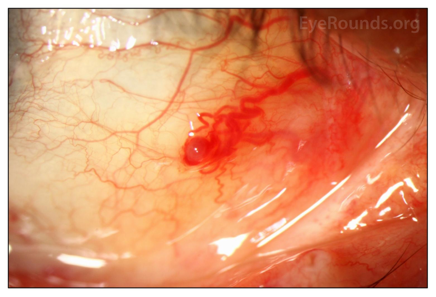
Slit lamp examination revealed a 1x1 mm superficial telangiectasia on the nasal bulbar conjunctiva OD (Figure 1). The bleeding was thought to be likely due to minor ocular surface trauma secondary to eye rubbing around the telangiectasia, resulting in the reported bloody tears. She has underlying dry eye disease and seasonal allergies that may have contributed to irritation and ocular surface fragility. Her baseline blurry vision was thought to be due to her dry eye symptoms.


DIAGNOSIS: Hemolacria secondary to Hereditary Hemorrhagic Telangiectasia


DISCUSSION

Hemolacria (bloody tears) is a rare condition with a broad differential diagnosis ranging from idiopathic, infectious, inflammatory, vascular, traumatic, and neoplastic etiologies to side effects of topical medications (e.g., silver nitrate) [1]. However, most cases of hemolacria are secondary to benign conjunctival lesions [2]. Due to the association between severe systemic and ocular diseases, a thorough evaluation is recommended for all patients presenting with new onset hemolacria. Patients should undergo a comprehensive ophthalmic and systemic examination to identify organic causes or sources of bleeding, whether it be from the conjunctiva, lacrimal puncta, lid margin, tear glands, or orbit [1]. Treatment of hemolacria is diverse and is aimed at correcting the underlying condition. If the history, physical exam, and imaging studies are consistent with a benign conjunctival lesion, it is reasonable to observe to ensure resolution of symptoms. If an infection is suspected, application of topical antibiotics may be appropriate.

Hereditary hemorrhagic telangiectasia (Rendu-Osler-Weber disease) is a rare genetic disorder characterized by the formation of vascular malformations. Diagnosis of HHT is based on Curaçao's clinical criteria, which include the presence of multiple telangiectasias in the skin and mucous membranes, recurrent epistaxis, visceral involvement (e.g., arteriovenous malformations in the lung, brain or liver) and a first degree relative with HHT [3]. It is inherited in an autosomal dominant fashion and is commonly caused by mutations in the ENG (i.e., HHT1 subtype) or ALK1 genes (i.e., HHT2 subtype). Ocular involvement is common in HHT and reports of hemolacria in HHT patients have been previously documented [4]. In a previous cross-sectional study of 206 HHT patients, 44.1% of patients harboring ALK1 mutations developed ocular telangiectasias [3]. Most ocular telangiectasias were located in the tarsal conjunctiva, bulbar conjunctiva, lid margin, and caruncle [3].

EPIDEMIOLOGY

  • Hemolacria is exceedingly rare. Thus, the predilection for a specific sex, age group, or race is unknown.

SIGNS

  • Conjunctival and/or scleral injection
  • Bloody, clear, and/or mucoid drainage
  • Presence of vascular or mass lesions on the conjunctiva or lacrimal apparatus

SYMPTOMS

  • Bloody drainage (tears)
  • Clear or mucoid drainage
  • Blurry vision
  • Pain if source of blood is traumatic, inflammatory, or infectious
  • Psychological distress

TREATMENT/MANAGEMENT

  • Diagnostic work up includes: Thorough slit lamp examination of the conjunctiva, lacrimal gland, and puncta for lesions. Additional diagnostic procedures may include probing and irrigation of lesions, culture or cytology of discharge from puncta, nasal endoscopy, CT scan of the sinuses, dacryocystography, or intraoperative biopsy.
  • Treat underlying systemic disease if such conditions are present. Observation and close follow-up with or without topical therapy (e.g., antibiotics in the case of suspected infection) may be most appropriate to ensure resolution of symptoms.

References

  • 1. Tripathy, K. and B. Salini, Hemolacria, in StatPearls. 2020: Treasure Island (FL).
  • 2. Murube, J., Bloody tears: historical review and report of a new case. Ocul Surf, 2011. 9(3): p. 117-25.
  • 3. Gomez-Acebo, I., et al., Ocular lesions in hereditary hemorrhagic telangiectasia: genetics and clinical characteristics. Orphanet J Rare Dis, 2020. 15(1): p. 168.
  • 4. Soong, H.K. and D.A. Pollock, Hereditary hemorrhagic telangiectasia diagnosed by the ophthalmologist. Cornea, 2000. 19(6): p. 849-50.

Suggested citation format:

Velez G, Dotson AD, Oetting TA. Hemolacria Secondary to Hereditary Hemorrhagic Telangiectasia. EyeRounds.org. June 18, 2021. Available from https://eyerounds.org/cases/313-Hereditary-Hemorrhagic-Telangiectasia.htm


Image Permissions:
Creative Commons

Ophthalmic Atlas Images by EyeRounds.org, The University of Iowa are licensed under a Creative Commons Attribution-NonCommercial-NoDerivs 3.0 Unported License.