INITIAL PRESENTATION
Chief Complaint: Blurred vision in the right eye for several weeks
History of Present Illness
A 70-year-old male presented with blurred vision in the right eye for several weeks. He was referred by a local optometrist for neovascularization of the iris and ocular hypertension in the right eye.
Past Ocular History
Past Medical History
Medications
Allergies
Family History
Social History
Review of Systems
OCULAR EXAMINATION
| OD | OS | |
|---|---|---|
| Lids/lashes | Normal | Normal |
| Conjunctiva/sclera | Complexion associated melanosis | Complexion associated melanosis |
| Cornea | 4+ punctate epithelial erosions (PEE) | 2+ PEE |
| Anterior Chamber | 2+ flare and rare cell | Deep and quiet |
| Iris | Superior peri-pupillary neovascularization of the iris | Normal architecture |
| Lens | Posterior chamber intraocular lens with anterior capsular phimosis | Posterior chamber intraocular lens with anterior capsular phimosis |
| Vitreous | Trace pigmented anterior vitreous cell; vitreous syneresis | No anterior vitreous cell; vitreous syneresis |
| OD | OS | |
|---|---|---|
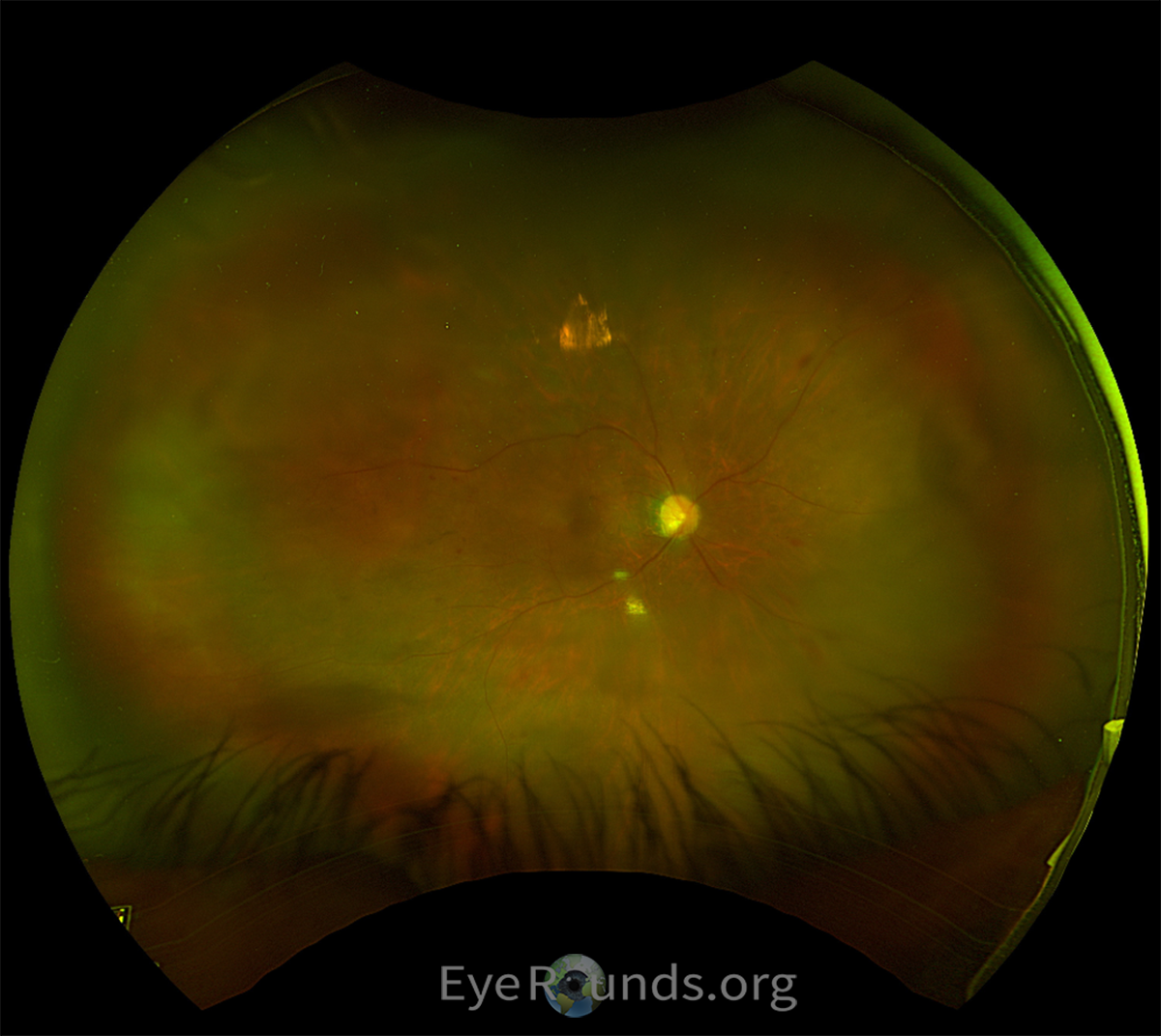
| Disc | Temporally tilted disc without disc hemorrhage; Inferior and superior thinning | Temporally tilted disc without disc hemorrhage |
| Cup-to-disc ratio | 0.8 | 0.6 |
| Macula | Moderate epiretinal membrane temporally, scattered hard drusen, many microaneurysms; Dot blot hemorrhages along arcades | Moderate epiretinal membrane temporally, scattered hard drusen, many microaneurysms |
| Vessels | Slightly attenuated | Slightly attenuated |
| Periphery | Numerous dot blot hemorrhages in mid-periphery (at least ¾ quadrants) | Dot blot hemorrhages were noted in 2/4 quadrants OS without mid-peripheral predilection. |
DIAGNOSIS: Ocular Ischemic Syndrome and secondary neovascular glaucoma in the right eye
CLINICAL COURSE
Based on right eye anterior chamber flare, mid-peripheral intra-retinal hemorrhages, and anterior segment neovascularization, the patient was diagnosed with neovascular glaucoma likely secondary to ocular ischemic syndrome. This diagnosis was complicated by the presence of pre-existing non-proliferative diabetic retinopathy bilaterally. The patient was given an intravitreal injection of the anti-VEGF medication Avastin (bevacizumab) in his right eye to control the anterior segment neovascularization. He had follow-up carotid ultrasound imaging, which did not reveal any significant stenosis. However, CT angiography of the head and neck showed prominent calcification within the ophthalmic branch of the internal carotid artery, which likely led to the development of ocular ischemic syndrome. Dorzolamide-timolol and prednisolone eye drops were added to patient’s medications to help control his intraocular pressure (IOP) and to help with pain. He continued to follow up monthly, and over time his vision continually improved. He eventually required surgical placement of a Baerveldt tube shunt in the right eye for IOP control and was counseled to continue glycemic and blood pressure control. Given that the area of stenosis was in the ophthalmic branch of the internal carotid artery rather than within the main portion of the internal carotid artery, no carotid endarterectomy or stenting was indicated.
DISCUSSION
Etiology/Pathophysiology
Ocular ischemic syndrome (OIS) consists of ocular hypoxia resulting from occlusion or stenosis of the common or internal carotid arteries [1]. Stenosis is typically severe, occluding approximately 90% or more of the common or internal carotid arteries or their branches ipsilateral to affected eye. Occlusion of the ophthalmic artery can also cause the development of OIS but is less common. The increased resistance in any of these vessels reduces blood flow and oxygen delivery to the eye as blood instead travels back towards the intracranial circuit rather than to the eye.
OIS occurs most frequently in the elderly population, with an average patient age of 65 years [1]. Cases rarely occur in individuals under 50 years of age. The incidence is twice as high in males compared to females; this may be due to higher rates of cardiovascular disease in males. Risk factors for OIS include cardiovascular disease, hypertension, and diabetes mellitus. Patients with OIS have as high as a 40% mortality rate within 5 years of onset, with mortality most often secondary to the associated cardiovascular disease [1].
Signs/Symptoms
In one study, OIS was found to cause decreased visual acuity in the affected eye in 90% of patients [2]. In 67% of these patients, the onset of visual loss occurred over weeks to months. Another 12% of patients experienced vision loss over the course of a few days, and a final 12% lost visual acuity over a matter of seconds to minutes. Patients with sudden vision loss often present with a cherry-red spot located over the fundus resulting from the embolization of the central retinal artery leading to a central retinal artery occlusion [2]. The visual fields of OIS patients vary based on the retinal pathology and secondary glaucoma. Additionally, a specific finding of OIS patients with severe carotid artery stenosis is that they will describe delayed recovery of visual function following exposure to bright light. On initial presentation, visual acuity ranges from 20/50 to 20/200 in 43% of patients, with 37% of patients’ vision diminished to counting fingers or worse. Vision typically declines over the subsequent 12 months, with 24% of patients at 20/50 to 20/200 and 58% at counting fingers or worse [1].
In addition to visual acuity and field deficits, 40% of patients with OIS present with pain in the affected eye most often due to elevated intraocular pressure (IOP) from secondary neovascular glaucoma or direct ischemia to the eye [2]. If the pain is due to ischemia, it typically develops gradually and presents as a constant, dull ache in the affected eye that radiates to the upper face and temple.
Anterior segment signs of OIS include neovascularization of both the iris and the iridocorneal angle, seen in 66% of patients, and neovascular glaucoma, which occurs in 50% of patients [1]. Less frequently, a mild iritis is seen – when this is observed, flare is more pronounced than cellular reaction due to ischemic damage [2,3]. In cases where neovascular glaucoma occurs, optic nerve damage may result [1]. Even in cases with neovascularization of the angle, the IOP may not be high due to poor perfusion of the ciliary body and low aqueous production. Additional anterior segment signs include iris atrophy and corneal edema (reflecting chronic ischemia) as well as asymmetric cataract [2].
Posterior signs are more frequently observed in eyes with OIS, such as dilated retinal veins and narrowed retinal arteries [1]. Intra-retinal hemorrhages are observed in most affected eyes because of retinal capillary damage leading to microaneurysm rupture or leakage of blood [1-3]. These hemorrhages are classically located in the mid-periphery and are almost never confluent.
Testing
Carotid artery imaging with Duplex carotid ultrasonography is a crucial aspect of diagnosing OIS – with imaging, 75% of carotid stenosis cases can be diagnosed non-invasively [1]. Imaging of retrobulbar vessels including the ophthalmic artery, the central retinal artery, and short posterior ciliary arteries can be performed if OIS is suspected despite healthy carotid arteries [1,2]. This is accomplished best with CTA rather than MRA due to the shortened imaging time needed to accomplish a CTA relative to an MRA. Fluorescein angiography of the fundus displays prolonged or irregular filling time of both the choroid and retinal circulation in 60% of patients with OIS [1]. Indocyanine green (ICG) angiography is rarely performed but can help confirm an OIS diagnosis by demonstrating the prolonged arm-to-choroid circulation time and intra-choroidal circulation time.
Differentiating OIS and Diabetic Retinopathy
OIS shares many similarities with diabetic retinopathy and the overlapping risk factors can make this diagnosis challenging. Our patient had a background of mild-to-moderate non-proliferative diabetic retinopathy; however, the asymmetry of the findings, mid-peripheral predilection of the intra-retinal hemorrhages, and marked anterior chamber flare helped to distinguish OIS from diabetic retinopathy alone.
Neovascularization of the iris and iridocorneal angle is seen in 66% of patients with OIS, leading to neovascular glaucoma in 50% of cases [1]. While neovascular glaucoma resulting from neovascularization of the iris can occur in diabetic retinopathy, many of these patients have posterior findings of proliferative diabetic retinopathy which were absent in this patient.
In unclear cases, diagnostic testing can help distinguish between diabetic retinopathy and OIS. Approximately 20% of patients with diabetic retinopathy have hemodynamically significant carotid artery stenosis [1]. As such, significant asymmetry of retinopathy should prompt imaging to evaluate for carotid artery stenosis. While fluorescein angiography in patients with OIS often shows prolonged or irregular filling time of choroid and retinal circulation, filling times should be normal in diabetic retinopathy alone.
Treatment
Primary treatment of OIS is treatment of the stenosed vessel [1,2]. Carotid artery endarterectomy (CEA) is used to treat carotid artery stenosis that is 99% or less stenosed with an efficacy of 70-90%. Carotid artery stenting can also be utilized in cases without complete arterial occlusion. Both techniques help restore blood flow to the affected eye with a goal of improving perfusion and reversing some of the effects of OIS. If the artery is completely (100%) occluded, surgery is avoided because either there are collaterals already at work or the downstream ischemic damage is already done.
Once reperfusion is established, visual acuity can be stabilized or restored, especially if restorative procedures are performed before neovascularization and glaucoma occur [1]. Both neovascularization and glaucoma can be reduced if bypass surgery is performed in the early stages of neovascular glaucoma. However, once reperfusion has occurred, there is often a marked increase in IOP because of the restoration of aqueous humor production.
Management of ischemic complications is also often needed for better visual outcomes. For patients with neovascularization, laser pan-retinal photocoagulation is used to ablate the non-functional peripheral retina and decrease retinal ischemic drive. Intravitreal anti-VEGF medications can also be used to decrease the presence of intraocular VEGF molecules that drive the process of neovascularization [1]. If neovascular glaucoma is present, surgical management with a tube shunt is often necessary. If the IOP is very high, a valved tube shunt may be required, although occasionally the IOP is low enough to wait for a non-valved tube shunt to open. Trabeculectomies often fail in neovascular glaucoma cases due to significant bleeding. Although it may be reasonable to temporize with medical management with IOP lowering drops, this is often not sufficient to control the IOP if the angle is completely closed.
DIFFERENTIAL DIAGNOSIS:
EPIDEMIOLOGY OR ETIOLOGY
|
SIGNS
|
SYMPTOMS
|
TREATMENT/MANAGEMENT
|
References
Shepard ZV, Hendricks TM, Boese EA. Ocular Ischemic Syndrome in a patient with background Diabetic Retinopathy: Distinguishing Features. EyeRounds.org. March 21, 2024. Available from https://eyerounds.org/cases/356-ocular-ischemic-syndrome.htm

Ophthalmic Atlas Images by EyeRounds.org, The University of Iowa are licensed under a Creative Commons Attribution-NonCommercial-NoDerivs 3.0 Unported License.

Address
University of IowaLegal
Related Links