
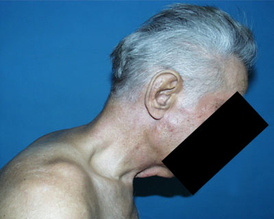
Case Presentation: A 65–year-old male with a visually significant cataract presented to our clinic with severe kyphosis caused by ankylosing spondylitis (Figure 1). Kyposis severely limited his neck extension; he was unable assume the typical supine position we use for cataract surgery.
 |
Discussion: Kyphosis is curvature of the spine causing a "hunchback" or roundback. It is often a congenital disease but may also be caused by osteoporosis, degenerative arthritis of the spine, trauma, developmental problems, infectious disease of the spine, or connective tissue disorders. Kyphosis is a rare complication of ankylosing spondylitis. Ankylosing spondylitis is a chronic autoimmune condition associated with antigen HLA B27. Uveitis, heart valve disease, inflammation of the aorta, and pulmonary fibrosis can also develop.
Positioning patients with kyphosis presents a very difficult challenge for the cataract surgeon. One approach is to lay the patient in the usual position and operate with the head more vertical than usual. This makes the operation quite different for the surgeon. Dr. Mark Packer has described his approach to operating on eyes in this more vertical position in an informative video. (http://www.surgytec.com/video/cataract-surgery-kyphotic-patient).
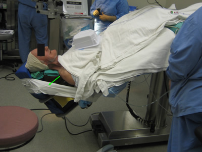
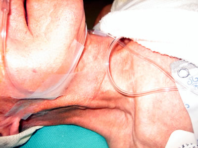
Course: Our approach was similar to that described by Gordon et al (2). Specifically, we raised the patient’s legs, which allowed the patient’s head to be flat. Using several foam devices, we created a large firm pillow, which we positioned between the head of the bed and the patient’s head, neck, and shoulders. While we were concerned about the risk of posterior pressure and noted some jugular venous distension (Figure 3), this did not cause difficulties during the surgery.
 |
 |
Conclusion: Positioning the patient in this particular way allowed us to perform the surgery in the usual fashion rather than vertically which made us more comfortable proceeding. The patient reported no discomfort during or after the procedure.
Jensen LE, Muthialu A, Oetting TA. Kyphotic Patient Presents for Cataract Surgery: A Systems Based Case. EyeRounds.org. December 8, 2008; Available from: http://www.EyeRounds.org/cases/90-Positioning-Patient-Kyphosis-Systems.htm.

Ophthalmic Atlas Images by EyeRounds.org, The University of Iowa are licensed under a Creative Commons Attribution-NonCommercial-NoDerivs 3.0 Unported License.

Address
University of IowaLegal
Related Links
