Chief Complaint: 4-year-old child with a history of seizures and glaucoma.
History of Present Illness: Patient was born with a reddish color to the face that respects the midline in the trigeminal nerve distribution. He also developed elevated intraocular pressures on the same side of the face lesion.
PMH: As stated above. FH: No previous family history of ocular diseases. POH: As stated above.
EXAM
| OD | OS |
 |
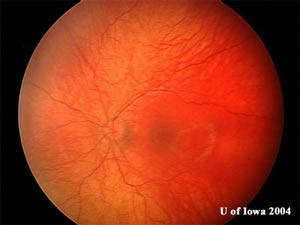 |
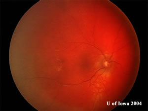
| Choroidal hemangioma with tomato catsup appearance of the fundus obscuring choroidal details. | Normal appearing fundus.
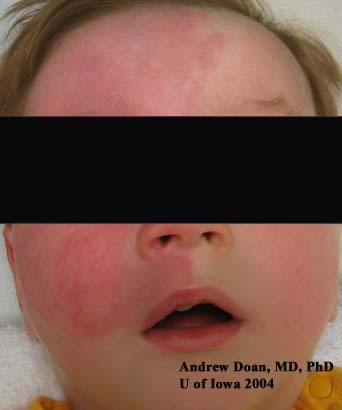 |
| Port-wine stain (facial hemangioma), a.k.a. nevus flammeus, of the right side of the face respecting the mid-line and in the distribution of the trigeminal nerve. |
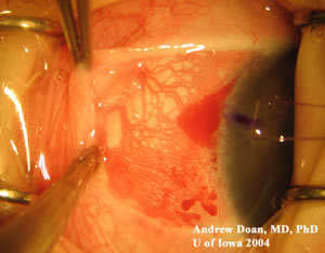
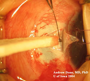
Our patient underwent trabeculotomy surgery as a baby to reduce the intraocular pressures OD. These photos are of another patient who underwent the same procedure. Young Kwon, M.D., Ph.D. (Glaucoma Service) was the primary surgeon in this case.
| Episcleral Vessels | Scleral Flap |
 |
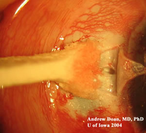 |
| The conjunctiva is pulled back revealing the dilated episcleral vessels due to increased episcleral venous pressure. | A scleral flap is made and dissection identifies Schlemm's canal. |
 |
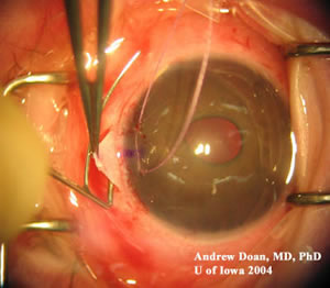 |
| 6-O Nylon suture is threaded into Schlemm's canal for confirmation. | One arm of the Harms trabeculotome is inserted into Schlemm's canal. |
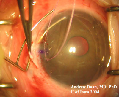 |
| A gentle twisting motion of the Harms trabeculotome allows entry into the anterior chamber. This motion allows formation of direct drainage of aqueous into Schlemm's canal. |
This is one of the phakomatoses (mother-spot disease) that is not inherited. The hamartomatous hemangiomas can be found on the face, eyelid (increases risk of glaucoma), choroid, and brain. These lesions are not malignant, but will cause local problems leading to glaucoma and seizures.
50% of patients will develop glaucoma, and 80% of patients may develop seizures. This disorder is rarely bilateral.
Pathophysiology: Resistance to aqueous outflow due to elevated episcleral venous pressure vs. underlying angle anomaly. Po=(F/C) + Pv [Goldmann Equation: Po = intraocular pressure in mmHg; F = rate of aqueous production in ml/min; C= facility of outflow in ml/min/mmHg; Pv = episcleral venous pressure in mmHg). Some believe there is a component of both elevated episcleral venous pressure and underlying angle anomaly contributing to increased intraocular pressures. Thus, patients may be managed with surgical trabeculotomy or goniotomy.
EPIDEMIOLOGY
|
SIGNS
|
SYMPTOMS
|
TREATMENT
|
Doan A, Kwon YH: Sturge-Weber Syndrome: 4-year-old child with a history of seizures and glaucoma. February 21, 2005; Available from: http://www.EyeRounds.org/cases/case13.htm.

Ophthalmic Atlas Images by EyeRounds.org, The University of Iowa are licensed under a Creative Commons Attribution-NonCommercial-NoDerivs 3.0 Unported License.