This site uses tracking information. Visit our privacy policy. Click to agree to this policy and not see this again.

Ophthalmology and Visual Sciences

Orbital Lymphoma:

60-year-old male who was evaluated by internal medicine 1 week earlier for "conjunctivitis" in the left eye

Orbital Lymphoma:

60-year-old male who was evaluated by internal medicine 1 week earlier for "conjunctivitis" in the left eye
Andrew Doan, MD, PhD, Thomas Oetting, MD
February 21, 2005

Chief Complaint: 60-year-old male who was evaluated 1 wk ago for "conjunctivitis" OS by internal medicine.

History of Present Illness: Patient complained of a painless, gradual redness of his left eye over the last several months. He does not report any pain, discharge, discomfort, or itching. He presented to the on-call ophthalmologist for evaluation. He pointed to some fullness of the superior aspect of the medial canthus OS with mild conjunctival injection. Pt also complained that things were "blurry" with a slight double image, which resolved when one eye was closed separately.

PMH: h/o non-Hodkin's lymphoma and in remission for numerous years. No other ocular history or medical problems. No h/o of ocular traumas or surgeries.

EXAM

  • Best corrected visual acuities: 20/20 OU.
  • Pupils: 4.5->3.5, no RAPD
  • EOM: elevation deficit OS with left hypotropia in primary gaze; VF Full to FC OU
  • IOP: 18 mmHg OD, 19 mmHg OS
  • DFE: retina exam notable for normal macula, vessels, and periphery OU. Optic nerves, normal.
  • SLE: OD normal, OS see photos below.
  • Hertel (base 113): 13 mm OD and 17 mm OS
Figure 1: EOM- Note the left hypotropia in primary gaze and elevation deficit OS with upgaze.
EOM

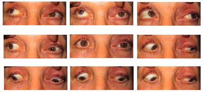
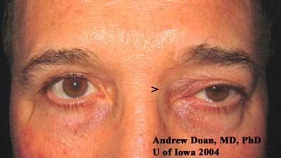
Figure 2: Left eye with superior conjunctival injection.
OS

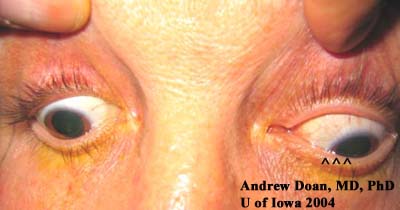
Figure 3: Left medial canthus with fullness. Note the asymmetric ptosis OS (more ptosis medially).
Ptosis

Figure 4: CT Scan

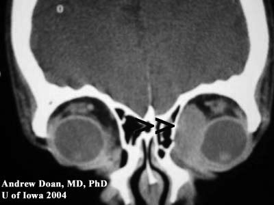
Coronal CT without contrast.

Axial CT without contrast.

Note: large medial mass indenting globe Note: large medial mass displacing medial rectus but is not eroding into bone or surrounding soft tissue.
CT CT

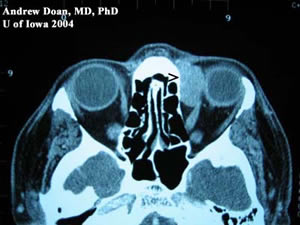
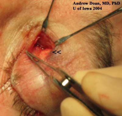
Figure 5: Anterior orbitotomy. Note the fleshy mass in the superior-nasal quadrant.
Anterior Orbitotomy

Discussion

Orbital Lymphoma

This patient presented with acute awareness of a long-standing problem: fullness of upper lid, binocular diplopia, and injection OS. He was diagnosed previously with "conjunctivitis" because of the asymmetric injection OS; however, he did not have other signs of infection: pain, itching, tearing, or discharge. On exam, his asymmetric ptosis OS (i.e. mechanical ptosis), elevation deficit OS on versions, proptosis OS, and a firm palpable mass in the superior-nasal quadrant OS should raise the suspicion of an orbital mass. In a patient with a history of non-Hodkin's lymphoma, orbital spread needs to be ruled out. The CT scans clearly denote a large orbital mass that is molding surrounding structures but not eroding into them.

An anterior orbitotomy was notable for a large, pink mass. Pathology confirmed the mass to be positive CD20 and BCL-2 lymphoma. Extensive imaging studies of head and body did not reveal additional tumor. The patient was treated by radiation oncology with local radiation.

Diagnosis: Orbital Lymphoma

EPIDEMIOLOGY

  • Incidence: in Florida, 2 cases per million
  • Age: all ages
  • Among the most common orbital tumors seen in the adults: cavernous hemangioma, lymphoid tumors, and meningiomas

SIGNS

  • Salmon patch lesion on globe
  • CT scan will show a lesion that pushes and molds surrounding structures and bone. There is no erosion into surrounding tissue.
  • Non-tender, firm mass

SYMPTOMS

  • Gradual onset
  • Painless
  • Slow progression
  • Binocular diplopia
  • Extraocular motility problems
  • Proptosis
  • Mechanical ptosis from tumor pushing down lid

TREATMENT

  • Depends on type of tumor and extension.
  • Chest and abdominal CT are used to identify abnormal nodes and spread.
  • Bone marrow aspiration to rule out marrow involvment.
  • If there is no systemic involvement, orbital lymphomas are typically sensitive to local radiation.
  • Chemotherapy is an option for patients with systemic involvment.
  • Incisional biopsy is needed for pathologic diagnosis.

Differential Diagnoses

  • lymphoid lesions of the orbit (benign reactive lymphoid hyperplasia, lymphoma, atypical lymphoid hyperplasia)
  • cavernous hemangioma
  • optic nerve meningioma
  • orbital metastasis
  • neurofibroma
  • neurilemoma (a.k.a. benign schwannoma)
  • fibrous histiocytoma
  • hemangiopericytoma
  • lymphangioma
  • mucocele
  • thyroid disease (most common cause of unilateral proptosis in adults)

References
  1. Hasegawa M, Kojima M, Shioya M, Tamaki Y, Saitoh J, Sakurai H, Kitamoto Y, Suzuki Y, Niibe H, Nakano T. Treatment results of radiotherapy for malignant lymphoma of the orbit and histopathologic review according to the WHO classification.Int J Radiat Oncol Biol Phys. 2003 Sep 1;57(1):172-6.
  2. Margo CE, Mulla ZD. Malignant tumors of the orbit. Analysis of the Florida Cancer Registry.Ophthalmology. 1998 Jan;105(1):185-90.
  3. Nerad, JA. Oculoplastic Surgery. The Requisites in Ophthalmology. Mosby 2001. 348-386.
Suggested citation format:

Doan A, Oetting TA: Verticillata: Orbital Lymphoma: 60-year-old male who was evaluated 1 wk ago for "conjunctivitis" OS by internal medicine. February 21, 2005; Available from: http://www.EyeRounds.org/cases/case9.htm.


last updated: 02-01-2005

Image Permissions:
Creative Commons

Ophthalmic Atlas Images by EyeRounds.org, The University of Iowa are licensed under a Creative Commons Attribution-NonCommercial-NoDerivs 3.0 Unported License.