University of Iowa Health Care

Ophthalmology and Visual Sciences

EyeRounds.org

EyeRounds Photo Quiz #1

Compiled by: Tyler Risma, M4

Click "Photo #" to see higher resolution, attempt to answer all questions related to a photo before clicking to see the answer.

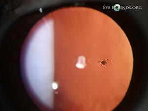
Photo 1 see enlarged


Q1: What is the diagnosis?

Q2:  What is the classic clinical appearance of the infiltrate in this condition?

Q3:  What imaging modality can help make the diagnosis?

Acanthamoeba keratitisAcanthamoeba keratitis


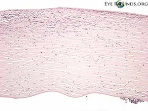
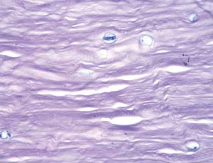
Photo 2 see enlarged

Q1: What is the differential diagnosis for this finding?

right eye, Amiodarone vortex keratopathyleft eye, Amiodarone vortex keratopathy


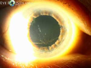
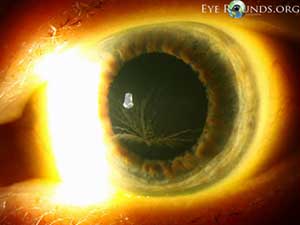
Photo 3 see enlarged

Q1: What are the two most important findings on this gonioscopic image?

Q2: What is the likely etiology and pathologic process?

Angle recession and cyclodialysis


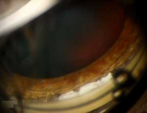
Photo 4 see enlarged

Q1: What are 3 important clinical findings in this photo?

Q2: What is the likely diagnosis?

Q3: What other ocular and systemic findings might be present?

Axenfeld-Rieger Syndrome


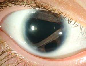
Photo 5 see enlarged

Q1: What is the abnormal finding in this histologic slide?

Q2: What is the diagnosis?

Band Keratopathy


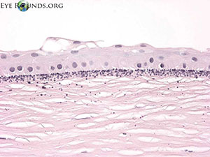
Photo 6 see enlarged

Q1: What clinical findings are present?

Q2: A 6 year old female with previously normal vision presents with this fundus appearance and worsening vision over the past 6 months to 20/200. What diagnosis must be on your differential, and a deletion in what gene is responsible?

Q3: What would an electroretinogram (ERG) likely show?

Right eyeLeft Eye


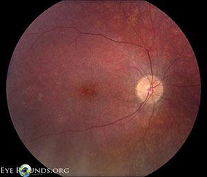
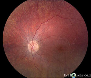
Photo 7 see enlarged

Q1: A 47 year old female with inflammatory cells in the anterior chamber and vitreous of both eyes presents with this fundus appearance. What is the most likely diagnosis?

Q2: What laboratory test would be most helpful to support the diagnosis?

Birdshot chorioretinopathy-right eyebirdshot chorioretinopathy

Birdshot choroiretinopathy, fluorescein angiographyBirdshot choroiretinopathy, fluorescein angiography


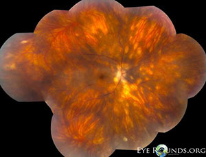
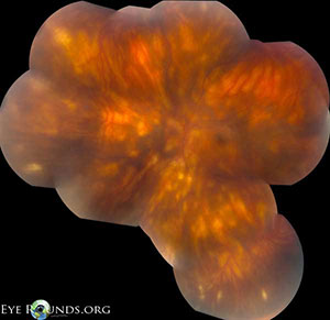
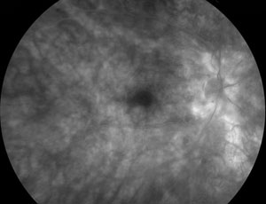
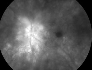
Photo 8 see enlarged

Q1: An 18-year-old male with this fundus appearance has a maternal uncle with poor vision. What is the most likely diagnosis and what gene is involved?

Choroideremia, right eyeChoroideremia, lefteye


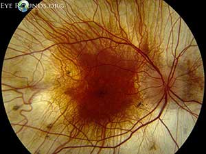
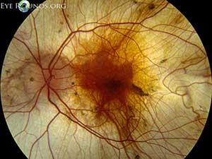
Photo 9 see enlarged

Q1: What symptoms might support the diagnosis of elevated intracranial pressure in this patient?

Q2:  How would you grade the papilledema?

Idiopathic Intracranial HypertensionIdiopathic Intracranial Hypertension

Idiopathic Intracranial HypertensionIdiopathic Intracranial Hypertension

Idiopathic Intracranial HypertensionIdiopathic Intracranial Hypertension

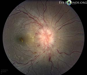
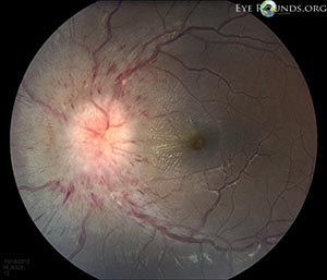
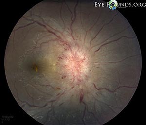
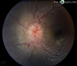
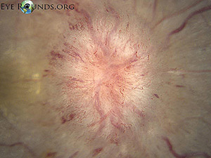
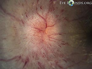
Photo 10 see enlarged

Q1: What is the diagnosis?

Q2: How is the diagnosis made?

Q3: What is the treatment?

Gyrate Atrophy, right eyeGyrate Atrophy


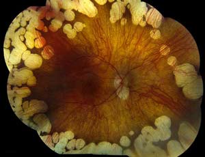
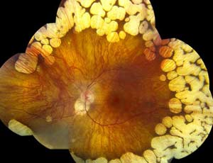
Photo 11 see enlarged

Q1: What is the name of this finding around the disc?

Q2: What is the cause?

High water mark after optic nerve head edema


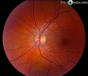
Photo 12 see enlarged

Q1: What are the pertinent findings in this photograph and what is the most likely etiology?

Iridodialysis and traumatic cataract after firecracker injury


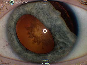
Photo 13 see enlarged

Q1: If a patient presented with a family history of this same ocular finding, what extraocular condition might you be concerned about?

Iris Flocculus


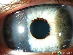
Photo 14 see enlarged

Q1: This finding reportedly occurs more frequently in patients with what type of glaucoma?

Optic disc hemorrahge in normal tension glaucoma


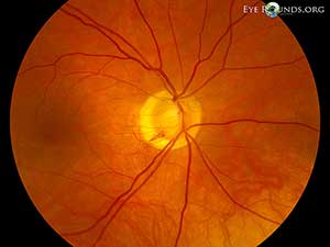
Photo 15 see enlarged

Q1: What type of visual field defect might you expect to see in this patient?

POAG, macula


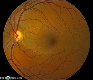
Photo 16 see enlarged

Q1: This 20-year-old male has a negative family history and 20/50 vision in both eyes. What is the most likely diagnosis?

Q2: How would you describe these yellow spots?

Stargardt Disease, right eyeStargardt Disease, left eye


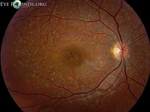
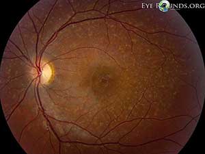
Photo 17 see enlarged

Q1: What are the pertinent findings in this slit lamp and gonioscopic photograph?

Q2:  In a patient with a history of trauma, what is the likely etiology?

Q3: What ocular problem is this patient at risk for?

Dilated Episcleral VesselsBlood in Schlemm's canal


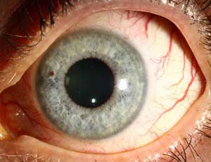
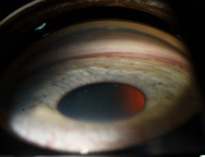
Photo 18 see enlarged

Q1: What is the name of this finding and where is it located?

Q2: What is the etiology?

Mittendorf dot

last updated: 9/29/2013
  Share this page: