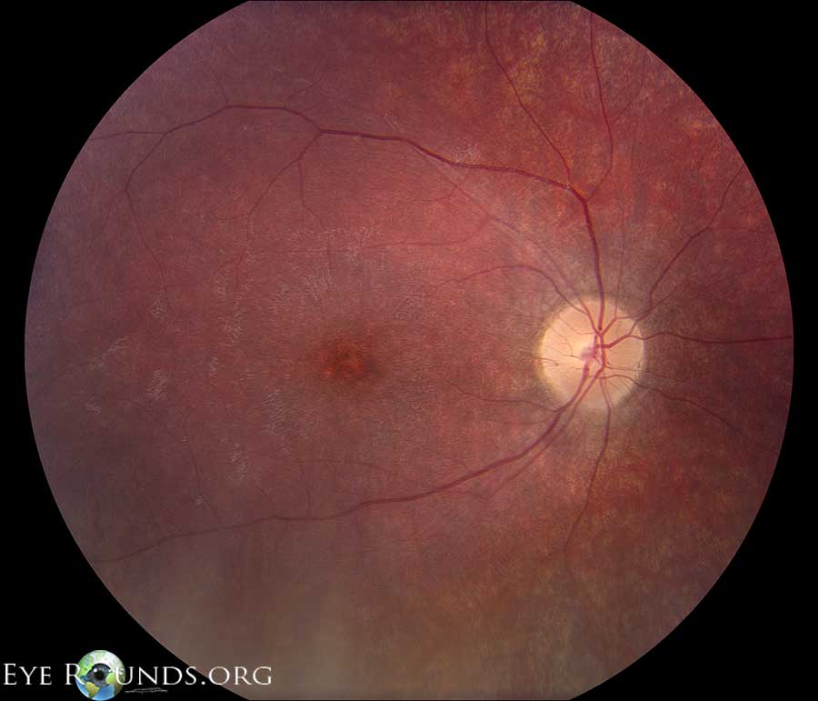
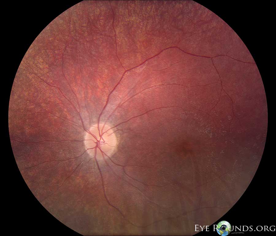
Photo 6
Q1: What clinical findings are present?
- mild optic nerve pallor, central macular mottling, attenuated arterioles, and peripheral retinal granularity
Q2: A 6 year old female with previously normal vision presents with this fundus appearance and worsening vision over the past 6 months to 20/200. What diagnosis must be on your differential, and a deletion in what gene is responsible?
- Batten disease (Juvenile Ceroid Lipofuscinosis); CNL3
Q3: What would an electroretinogram (ERG) likely show?
- The ERG would likely be non-recordable
see the Photo Atlas Entry← this link will open a new window, close it to continue the quiz
Right Eye
Left Eye


