This site uses tracking information. Visit our privacy policy. Click to agree to this policy and not see this again.

Ophthalmology and Visual Sciences

Idiopathic Intracranial Hypertension (IIH)

Idiopathic Intracranial Hypertension (IIH)

Category(ies): Neuro-ophthalmology
Contributor: Kimberly Winges, MD
Photographer: Toni Venckus, CRA

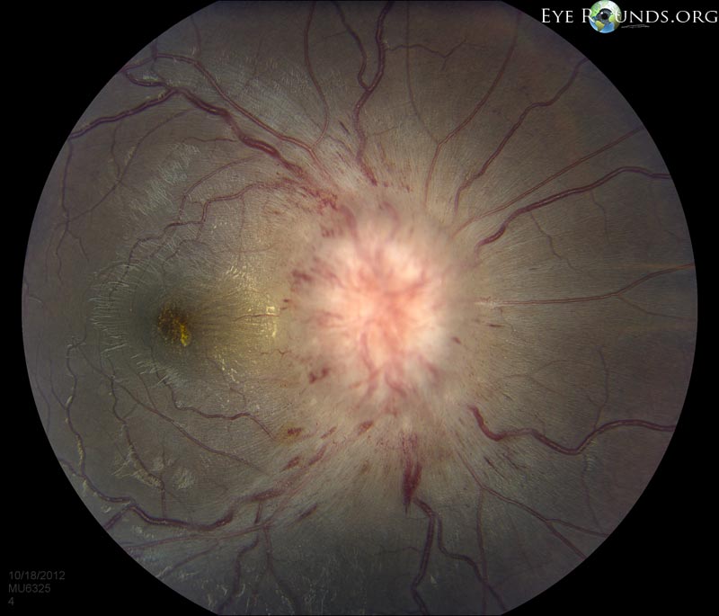
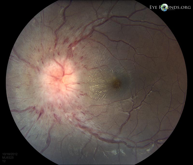
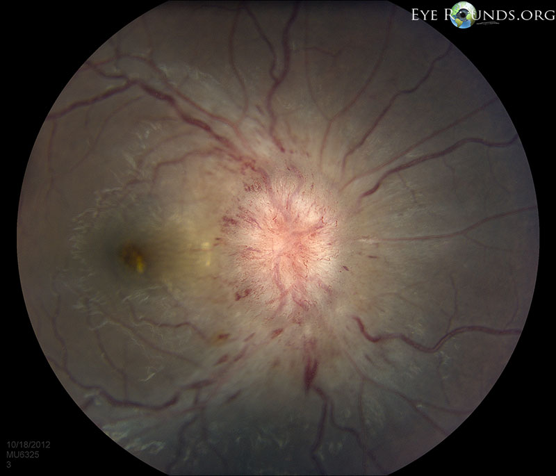
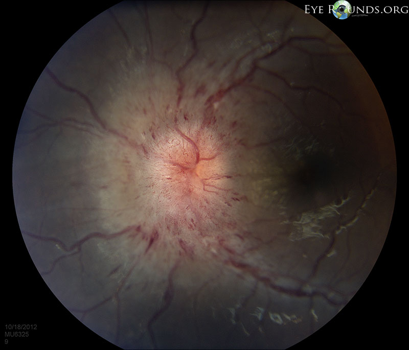
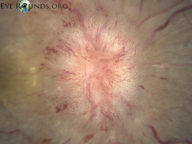
IIH first presentation in a 19-year-old Female with pulse-synchronous tinnitus and positional headache. Papilledema Grade 4 in both eyes.


Chronic papilledema from longstanding untreated IIH

Chronic papilledema from longstanding untreated IIH

Category(ies): Neuro-ophthalmology
Contributor: Lindsay Y. Chun MD; Sophia M. Chung MD
Photographer: Skiles, Sarah L
Posted: 07/01/2024

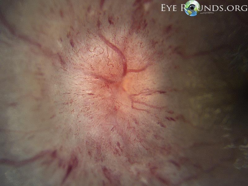
29 year-old female presenting with optic atrophy of the right eye and papilledema with scrolled glial veil of the left eye in setting of weight gain to over 300 pounds.

In the right eye photo there is severe optic nerve pallor associated with peripapillary fibrosis and perivascular changes related to severe and chronic disc edema.
In the left eye photo there is persistent disc edema with peripapillary changes, peripapillary wrinkles with an overlying glial veil, indicating persistent raised intracranial pressure.
In the right eye nerve OCT there is intraretinal schisis next to the right nerve and in the papillomacular bundle, and abrupt RPE atrophy.
In the left eye nerve OCT there is abrupt RPE atrophy as seen in the right eye.