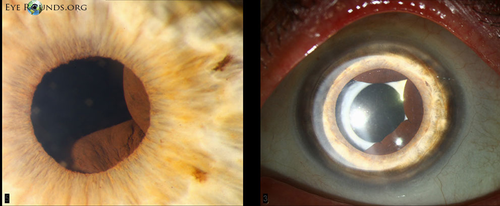
These cysts are fairly rare and classified as central (3%), midzonal (21%), peripheral (73%), and dislodged (3%). The majority of iris pigment epithelial cysts are benign and do not require any treatment.
Figure 1 The central cysts above were seen in a 62-year-old female with a history of angle closure glaucoma (perhaps due to enlargement of the cysts).
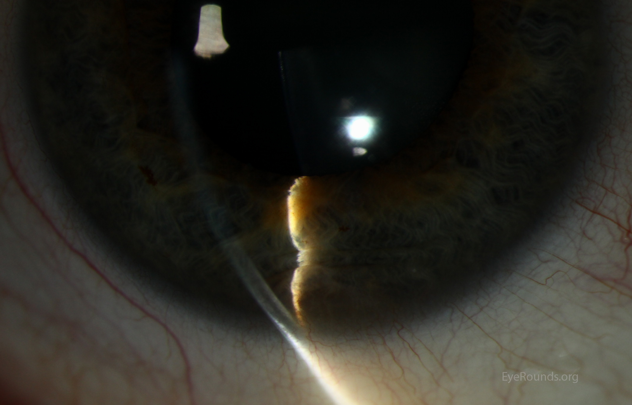
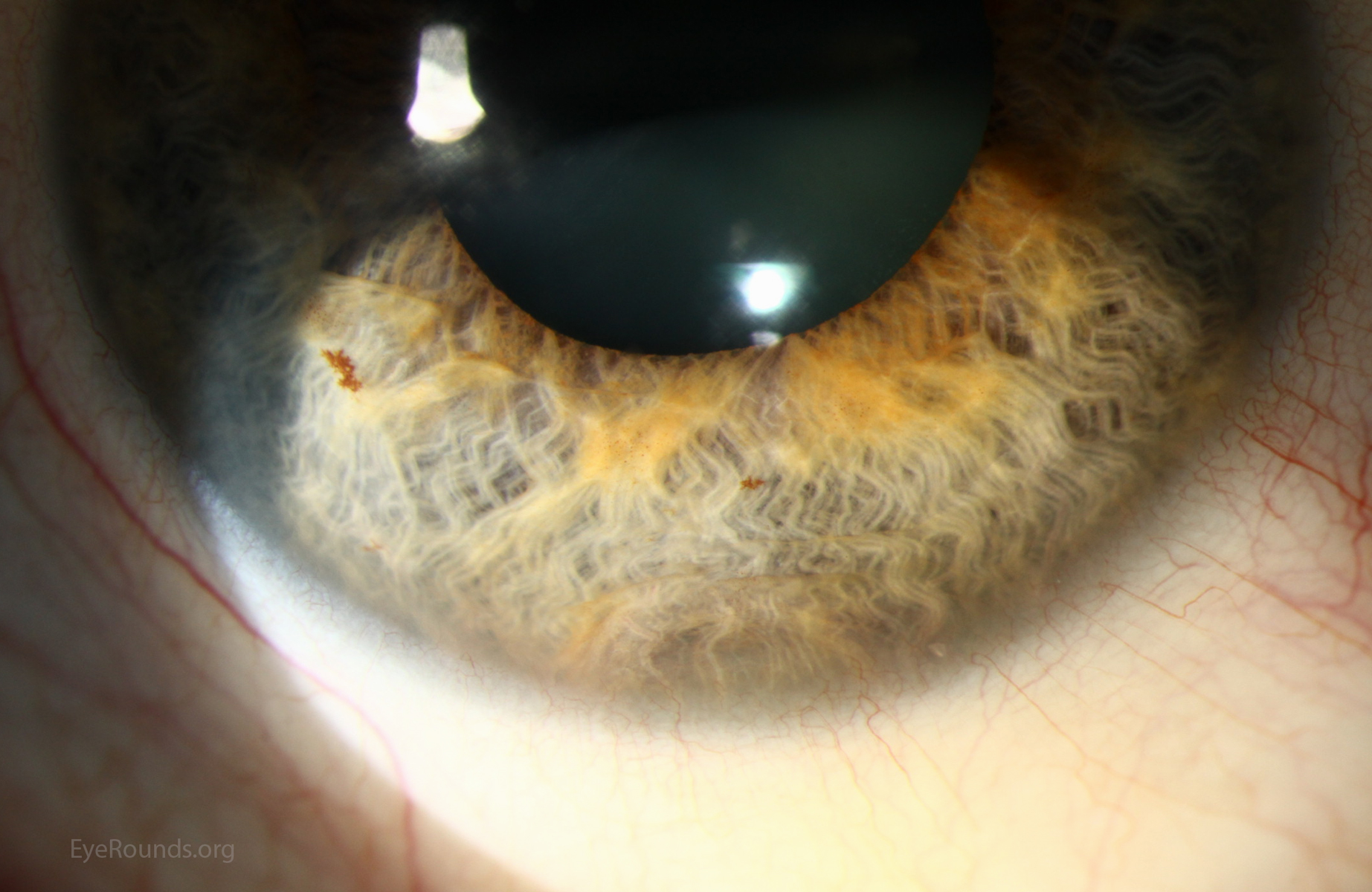
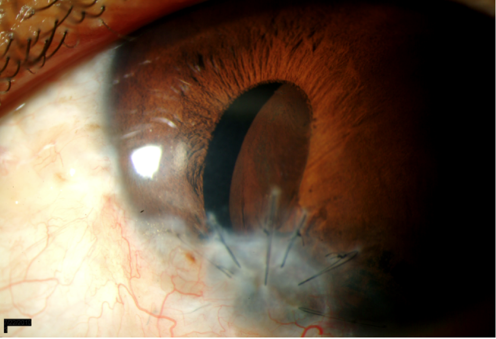
Figure 2A On slit lamp examination , an elevation of the inferior iris is seen. It appears as the iris is being pushed or bowed forward from a posterior mass. Folds in the iris can be seen.
Figure 2B On slit lamp examination , an elevation of the inferior iris is seen. It appears as the iris is being pushed or bowed forward from a posterior mass. Folds in the iris can be seen.
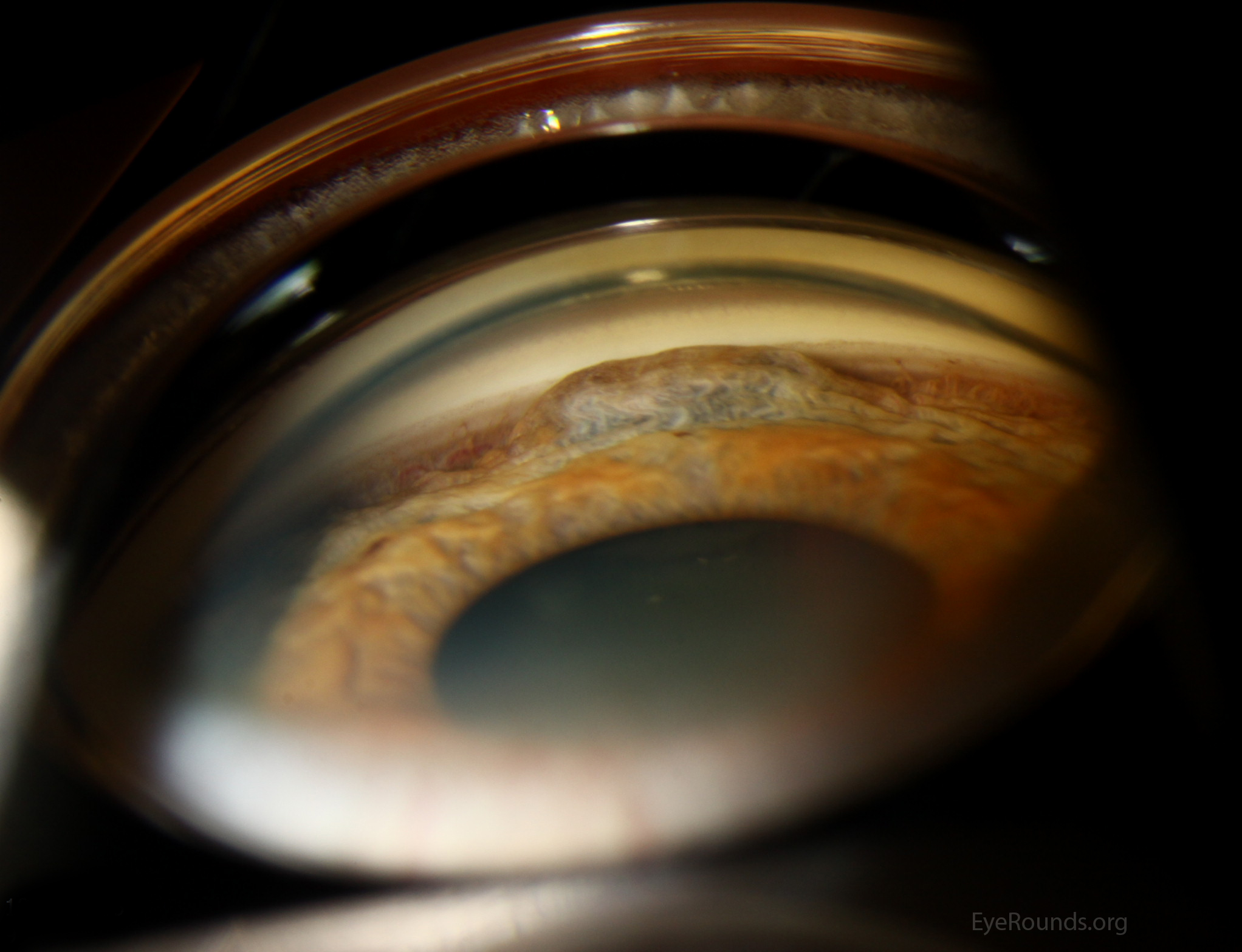
Figure 2C On gonioscopy examination (photo 2-C), the elevated, dome-shaped mass is apparent. With careful inspection, the pigment epithelial cyst can be seen through the pupil on the posterior surface of the iris. Also note the major arterial circle of the iris to the left of the cyst.
University of Iowa
Roy J. and Lucille A. Carver College of Medicine
Department of Ophthalmology and Visual Sciences
200 Hawkins Drive
Iowa City, IA 52242
University of Iowa
Roy J. and Lucille A. Carver College of Medicine
Department of Ophthalmology and Visual Sciences
200 Hawkins Drive
Iowa City, IA 52242