This site uses tracking information. Visit our privacy policy. Click to agree to this policy and not see this again.

Ophthalmology and Visual Sciences

Anterior Chamber Cilium

81-year-old male with intraocular cilium and dislocated intraocular lens

Anterior Chamber Cilium

81-year-old male with intraocular cilium and dislocated intraocular lens
Zachary Mayo, BS; Heather A. Stiff, MD; Prashant K. Parekh, MD, MBA; Jaclyn M. Haugsdal, MD; Thomas A. Oetting, MD, MS
posted July 5, 2017

INITIAL PRESENTATION

Chief Complaint

Referral for evaluation of dislocated intraocular lens (IOL) with posterior capsule opacification (PCO)

History of Present Illness           

An 81-year-old male with pseudophakia was referred to the University of Iowa Department of Ophthalmology for a dislocated IOL with PCO of the left eye. The patient had cataract extraction of the left eye with implantation of a 16.0 diopter 3- piece IOL (LI61AO) performed 17 months prior at an outside hospital. Per the surgical report, uncomplicated cataract extraction was performed with corneal cataract incision at 165 degrees and a corneal limbal relaxing incision at 340 degrees.

Four days following surgery, the patient was seen for follow up and no abnormalities were noted. In the following weeks the patient did not feel as if he was recovering as expected. He complained of floaters in his left eye ever since the surgery. Additionally, he experienced watering in his eye since surgery and often felt like "something was in the eye." He had developed shadows in his vision, which made it difficult to see and caused him to feel unstable at times. The patient felt that his vision was getting progressively worse. He denied any pain or redness in the left eye since the surgery.

Past Ocular History

Cataract extraction of both eyes with implantation of IOL
Myopia with astigmatism (-2.75 + 2.00 x 175 OS pre-surgery)
Left cataract post-surgical medications included

  • Combigan 0.2%-0.5% two times daily
  • Ketorolac 0.4% four times daily (QID)
  • Ofloxacin 0.3% QID
  • Prednisolone acetate 1% suspension QID

Past Medical History

Transient ischemic attack, abdominal aortic aneurysm, chronic obstructive pulmonary disease, hypercholesterolemia, enlarged prostate, acid reflux, cholecystectomy

Medications

Albuterol, amlodipine, aspirin, budesonide-formoterol, finasteride, montelukast, omeprazole, pravastatin, propylene glycol QID, tamsulosin

Allergies

None

Family History

History of cataracts in parent and sibling

Social History

Non-contributory

Review of Systems

Negative except for what is listed in HPI

OCULAR EXAMINATION

Current Spectacle Correction

  • Right Eye (OD): -0.25 + 1.50 x 004
  • Left Eye (OS): -0.25 + 0.75 x 180

Visual Acuity with correction (Snellen)

  • OD: 20/20
  • OS: 20/70 -2

Extraocular Motility

  • Both Eyes (OU): Full

Intraocular Pressure (IOP) Tonopen®

  • OD: 17 mmHg
  • OS: 15 mmHg

Pupils

  • OD: 3 mm (dark) → 2 mm (light) with no relative afferent pupillary defect (RAPD)
  • OS: 3 mm (dark) → 2 mm (light) with no RAPD

Confrontation visual fields with finger counting

  • OU: Full

External examination

  • OU: Normal

Slit lamp examination

  • OD
    • Lids/lashes: Meibomian gland dysfunction
    • Conjunctiva/sclera: Clear and quiet
    • Cornea: Clear
    • Anterior chamber: Deep and quiet
    • Iris: Normal Architecture
    • Lens: Posterior chamber intraocular lens centered and clear within the capsular bag with anterior capsular phimosis
  • OS
    • Lids/lashes: Meibomian gland dysfunction
    • Conjunctiva/sclera: Clear and quiet
    • Cornea: Nasal corneal wound
    • Anterior chamber: 6mm cilium with overlying pigment adherent to the iris at 6 o'clock just nasal to an area of iris atrophy (figure 1). The cilium extends towards the pupil. Anterior chamber is otherwise deep and without cell or flare
    • Iris: Areas of iris atrophy inferonasally and inferotemporally; peripupillary atrophy at 5 o'clock near intraocular cilium as noted above
    • Lens: 3-piece posterior chamber intraocular lens within the capsular bag (phimotic) displaced superiorly (figure 2); there is an anterior capsular tear inferiorly; posterior capsule intact with diffuse PCO; no pseudophacodonesis

Dilated Fundus Examination

  • OD
    • Vitreous: Normal
    • Optic nerve: Normal
    • Cup to disk (C/D) ratio: 0.3
    • Macula: Normal
    • Vessels: Normal
    • Periphery: Normal
  • OS
    • Vitreous: Normal - grossly
    • Optic nerve: Normal - grossly
    • C/D ratio: 0.3
    • Macula: Grossly flat
    • Vessels: Grossly normal
    • Periphery: Grossly attached

Additional Testing

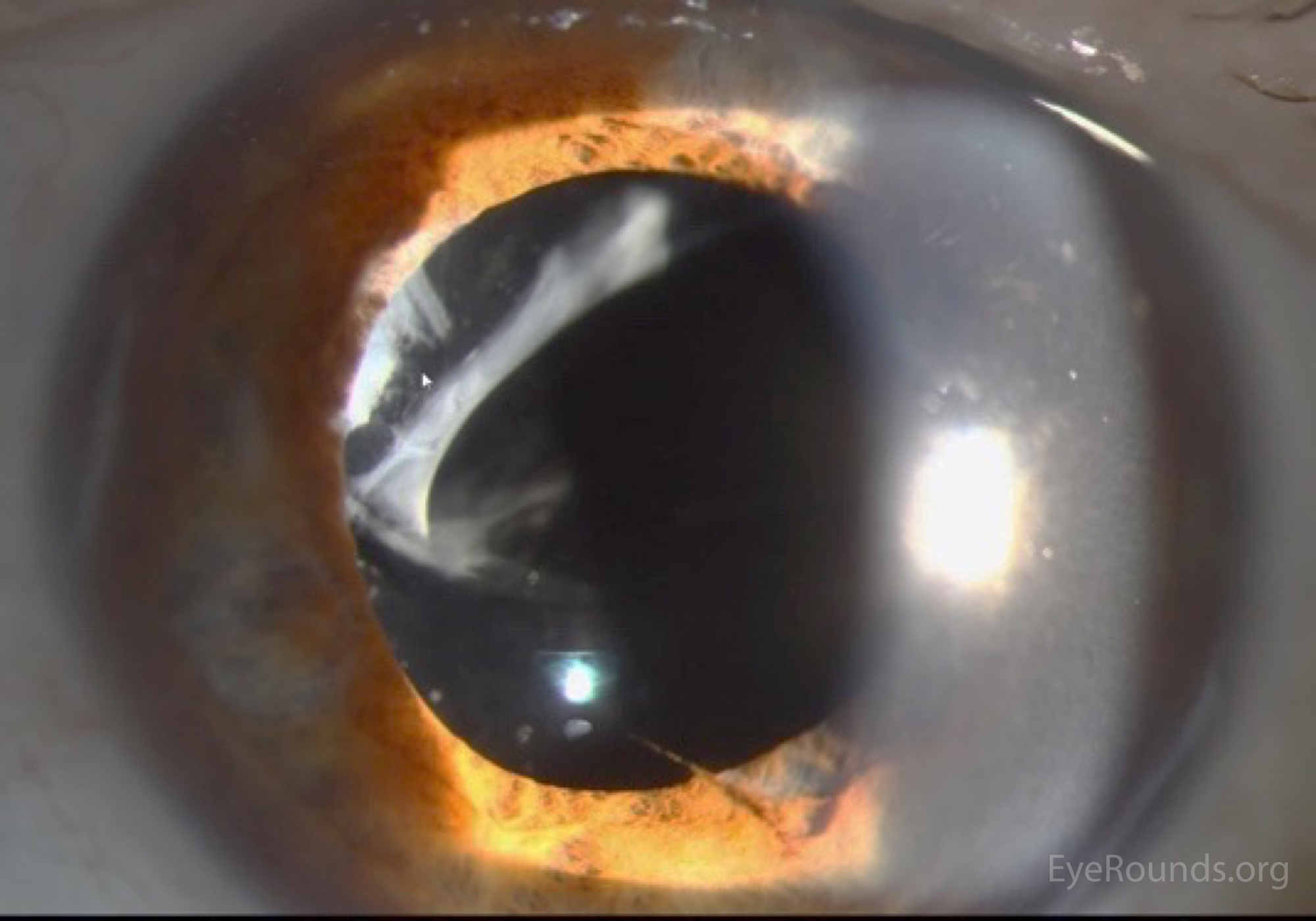
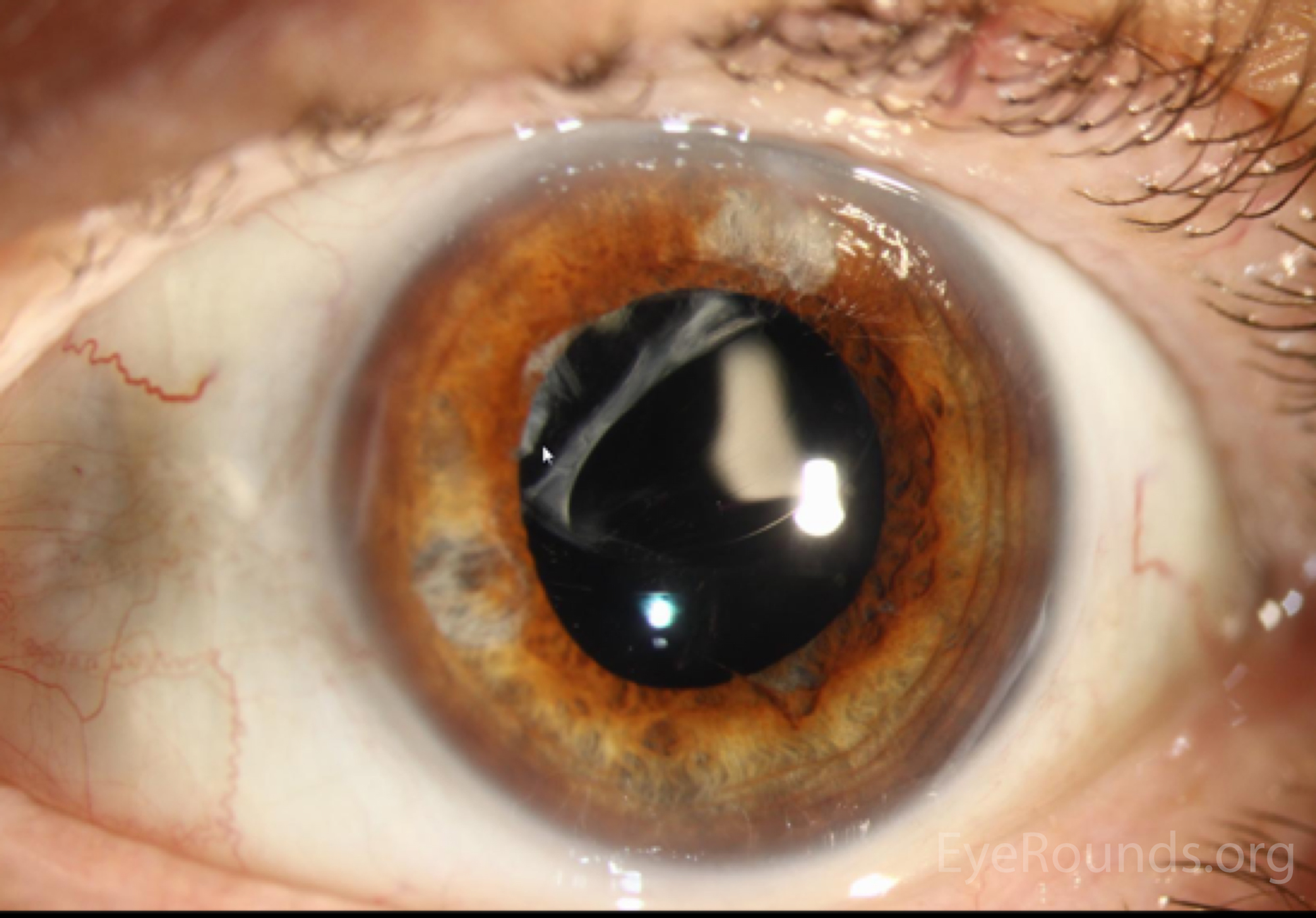
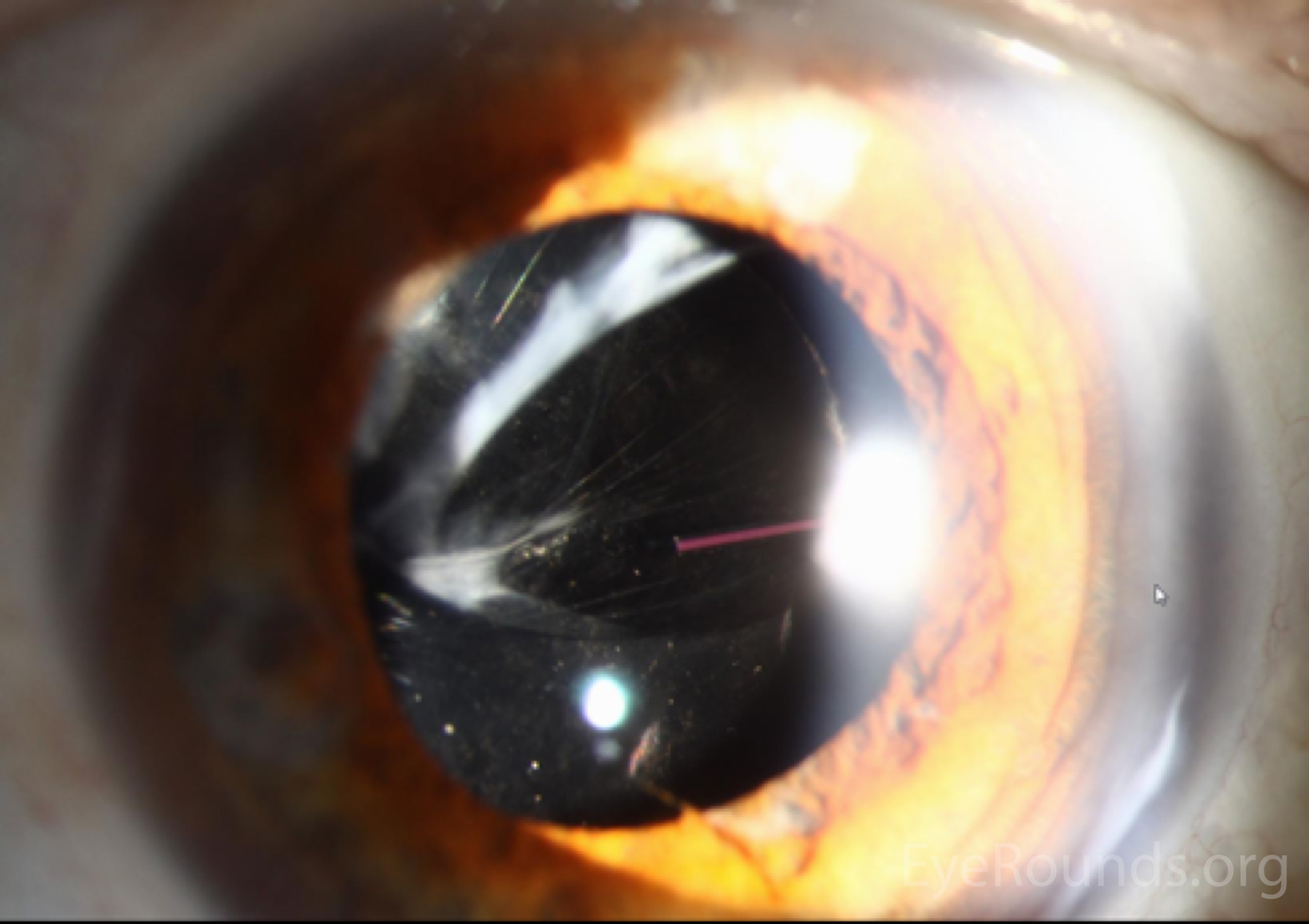
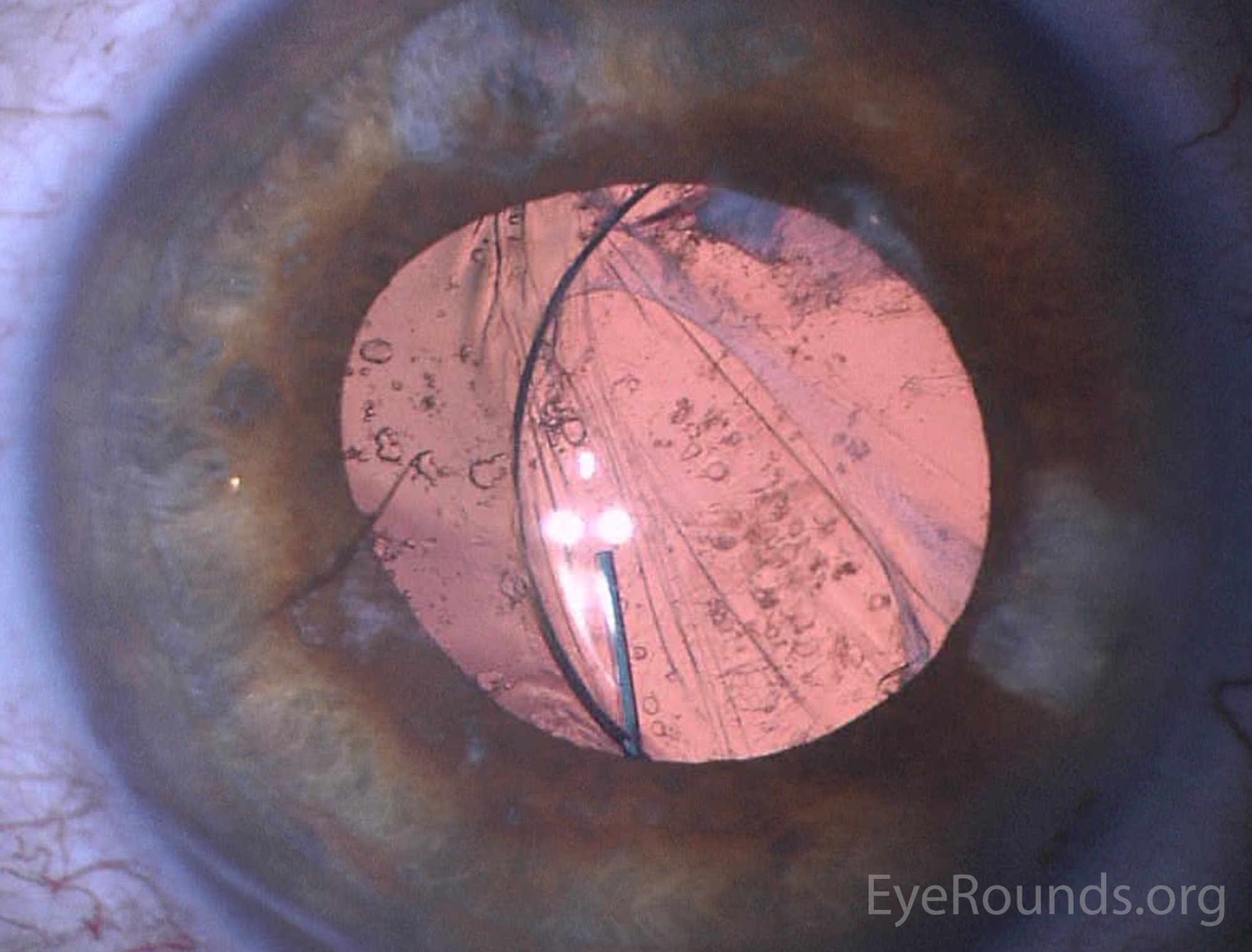
We obtained slit lamp photographs of the intraocular cilium (figure 1) as well as the dislocated intraocular lens (figure 2). We also obtained intraocular lens calculations.

Figure 1: Slit lamp photograph showing 6mm cilium with overlying pigment on the iris at 6 o'clock just nasal to an area of iris atrophy. The cilium extends towards the pupil.

Figure 1: Slit lamp photograph showing 6mm cilium with overlying pigment on the iris at 6 o'clock just nasal to an area of iris atrophy. The cilium extends towards the pupil.


Figure 2: Slit lamp photographs (A, B, and C) showing superior dislocation of a 3-piece posterior chamber intraocular lens within the capsular bag. Capsular phimosis is most notable superonasally in A and B. Retroillumination is used in C to show the dislocated lens, iris atrophy, and intraocular cilium at 6 o'clock. Also evident is the posterior capsule opacification.
Figure 2: Slit lamp photographs (A, B, and C) showing superior dislocation of a 3-piece posterior chamber intraocular lens within the capsular bag. Capsular phimosis is most notable superonasally in A and B. Retroillumination is used in C to show the dislocated lens, iris atrophy, and intraocular cilium at 6 o'clock. Also evident is the posterior capsule opacification. Figure 2: Slit lamp photographs (A, B, and C) showing superior dislocation of a 3-piece posterior chamber intraocular lens within the capsular bag. Capsular phimosis is most notable superonasally in A and B. Retroillumination is used in C to show the dislocated lens, iris atrophy, and intraocular cilium at 6 o'clock. Also evident is the posterior capsule opacification. Figure 2: Slit lamp photographs (A, B, and C) showing superior dislocation of a 3-piece posterior chamber intraocular lens within the capsular bag. Capsular phimosis is most notable superonasally in A and B. Retroillumination is used in C to show the dislocated lens, iris atrophy, and intraocular cilium at 6 o'clock. Also evident is the posterior capsule opacification.

Differential Diagnosis

  • Intraocular foreign body
  • Persistent pupillary strand/membrane
  • Iris suture
  • Fibrin in anterior chamber
  • Lysed posterior synechiae

CLINICAL COURSE

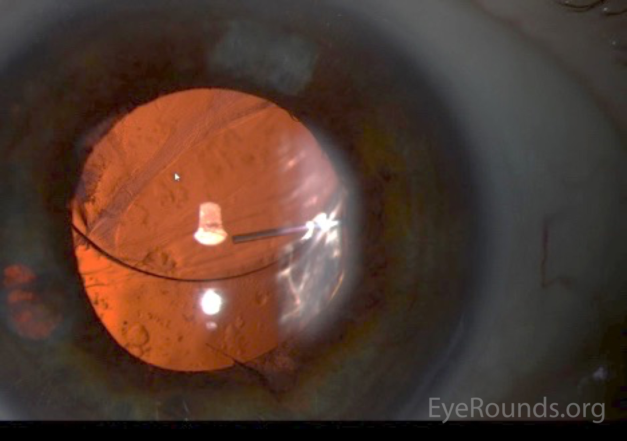
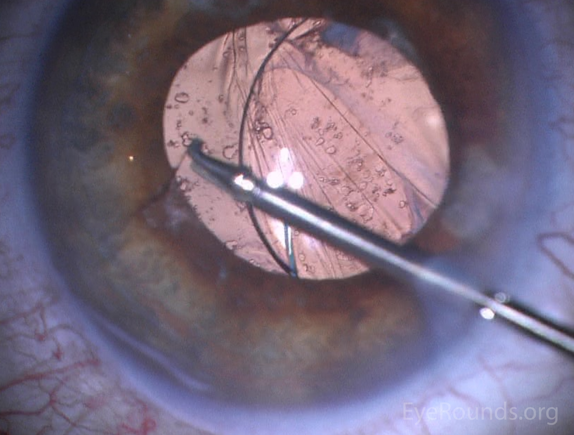
The patient was diagnosed with a dislocated IOL of the left eye with presence of an intraocular cilium. He was taken to the OR where the cilium was removed from the eye using duet forceps (figure 3, video). The existing 3-piece IOL was salvaged by placement into the ciliary sulcus with iris fixation. Post-operative slit lamp examination showed the IOL in good position.

Figure 3: Preoperative (3A) and intraoperative (3B) imaging showing removal of intraocular cilium with duet forceps.  (please see: Intraocular cilium removal following trauma)
Figure 3: Preoperative (3A) and intraoperative (3B) imaging showing removal of intraocular cilium with duet forceps.  (please see: Intraocular cilium removal following trauma) Figure 3: Preoperative (3A) and intraoperative (3B) imaging showing removal of intraocular cilium with duet forceps.  (please see: Intraocular cilium removal following trauma)

DIAGNOSIS

  • Visually significant dislocated intraocular lens of the left eye
  • Intraocular cilium located in the anterior chamber of the left eye

DISCUSSION

Etiology/Epidemiology

The finding of an anterior chamber cilium after small incision cataract surgery is rare, with only five prior cases being found on literature review (1-5). Intraocular cilia following ocular surgery (1-5) and ocular penetrating trauma (6-13) have been described in multiple locations of the eye, including the anterior chamber, posterior chamber, lens, vitreous, and retina (11).

Pathophysiology

Penetration of cilia into the eye is theoretically possible any time a patient has intraocular surgery. It is also speculated that inoculation may occur postoperatively as evidenced by observations of cilia penetrating through a corneal wound (3,4). This theory is supported by a case report of a patient who had a normal exam at 1 week after cataract surgery, but a cilium discovered in the anterior chamber at the 6 week follow up appointment (4). It is possible that eye rubbing may contribute to this process of post-operative inoculation (10). Cilium entering the eye due to trauma may occur by the penetrating object carrying the cilia into the eye or by an injury causing a blink reflex, and thereby forcing the cilium in (6-7). Video of intraocular cilium removal following trauma can be seen at
https://eyerounds.org/atlas-video/intraocular-cilium-removal-following-trauma.htm.

Cases of intraocular cilium in the absence of ocular trauma or prior ocular surgery have also been described (10,14,16). Kim and Henderson postulate that intraocular cilia may arise congenitally, as the surface ectoderm is the embryologic origin of both the cilia and the lens (16). Jin et al. report a case where they speculate a cilium migrated under the conjunctiva, leading to chronic bacterial infection and scleral necrosis. The cilium then migrated through the necrotic sclera to the vitreous cavity, resulting in bacterial endophthalmitis (14).

Signs/Symptoms

The response of the eye to cilia is unpredictable, ranging from severe intraocular inflammation (14) to a lack of reaction for 50 years (12). Reports have cited decreased visual acuity, foreign body sensation, bacterial endophthalmitis, plastic iridocyclitis, granulomatous inflammation, epithelial cysts, corneal decompensation, vitreous fibrosis, and retinal detachment as a result of intraocular cilium following surgery or trauma (6,8,10,14,15).

One case of cilium inoculation following small incision cataract surgery was reported by Galloway et al in 2004. In this case, the patient presented with anterior chamber cilium causing culture-negative-endophthalmitis 3 days after cataract surgery (1). Rofail et al. reported another case where an anterior chamber cilium was noted three days post-surgery. In this case the cilium pierced the corneal paracentesis, migrated into the posterior chamber, and re-emerged through the pupil to rest in the inferior angle. Six months later, this patient was still without any ocular inflammation (3). Islam and Dabbagh reported a third case where no inflammation was seen in the presence of an anterior chamber cilium for 4 years after cataract surgery (2).

Testing/Laboratory work-up/Imaging

The identification of an anterior chamber intraocular cilium is made with thorough slit lamp examination. If a cilium is discovered, slit lamp photos should be taken to follow the progress of the cilium, especially if it is not going to be removed. Seawright et al. reported that cilia in the posterior chamber can be detected with the use of B-scan ultrasonography. They reported the appearance of cilia on B-scan ultrasonography as curvilinear echogenic lesions with enhancement when the beam is at a right angle to the long axis of the cilium. This technique may help to distinguish cilia from calcific and metallic objects (13).

Treatment/Management/Guidelines

When considering the appropriate action to take after discovering an intraocular cilium, one must consider the benefits and risks of leaving the foreign body in the eye versus surgically removing it. Many reports have shown that intraocular cilia are inert, with the longest reported duration without complications being 50 years (12). However, there are cases that manifest serious complications such as uveitis and endophthalmitis (14). If no signs of inflammation or infection are seen, patients should be followed on an individual case-by-case basis, with consideration for removal at the earliest onset of inflammation or with suspicion for infection (2). Intervention in the absence of these signs should be considered if the cilium is located in a location that may damage the eye, such as the case where a cilium caused corneal endothelial cell deficiency (15), or in the case where two intraocular cilia led to retinal striations and disruption of macular anatomy (6).

Several precautions may be taken to avoid intraocular cilia during cataract surgery Authors of several case reports have given safety recommendations, such as having every individual in the operating room wear facemasks and caps/hairnets to minimize the risk of hair being shed into the surgical field (2). Additionally, surgical technique should focus on preventing eyelash exposure by using lid tape for accurate drape placement. If a foreign particle enters the eye during surgery, it should be flushed from the surgical zone immediately, before proceeding with the procedure (1). As multiple authors have proposed that cilia may enter the eye postoperatively, patients should be educated on the importance of avoiding eye-rubbing while the wound is healing (3,4).

Summary

Anterior Chamber Cilium

EPIDEMIOLOGY OR ETIOLOGY
  • History of corneal perforation, penetrating ocular trauma, or ocular surgery (1-13)
  • Rare during or after phacoemulsification procedure (1-5)
  • Non-traumatic inoculation (10,14,16)
  • Congenital (16)
SIGNS
  • Visualization of intraocular cilia with otherwise normal slit lamp exam
  • Adjacent corneal wound or scar
  • Associated corneal edema
  • Anterior chamber cell and flare
  • Epithelial cyst of iris or angle
  • Inflammation/infection of eye with unknown cause
SYMPTOMS
  • Often asymptomatic
  • Eye pain
  • Eye redness
  • Foreign body sensation
  • Decreased vision
  • Floaters


TREATMENT/MANAGEMENT
  • Asymptomatic: some choose to observe (2,3,5) but sometimes cilia are removed anyway, especially if easily accessible (4,6)
  • Signs/symptoms of infection, inflammation, or damage to surrounding structures: surgical removal (1,7,11,15)
References
  1. Galloway GD, Ang GS, Shenoy R, Beigi B. Retained anterior chamber cilium causing endophthalmitis after phacoemulsification. J Cataract Refract Surg 2004;30(2):521-522.  https://pubmed.gov/15030854
  2. Islam N, Dabbagh A. Inert intraocular eyelash foreign body following phacoemulsification cataract surgery. Acta Ophthalmol Scand 2006;84(3):432-434.  https://pubmed.gov/16704714
  3. Rofail M, Briner AM, Lee GA. Migratory intraocular cilium following phacoemulsification. Clin Exp Ophthalmol 2006;34(1):78-80.  https://pubmed.gov/16451264
  4. Walker NJ, Hann JV, Talbot AW. Postoperative cilium entrapment by clear corneal incision. J Cataract Refract Surg 2007;33(4):733-734.  https://pubmed.gov/17397752
  5. Bach A, Gold AS, Villegas VM, Wildner AC, Murray TG. Intraocular eyelash. J Am Osteopath Assoc 2015;115(2):115.  https://pubmed.gov/25637618
  6. Gupta AK, Ghosh B, Mazumdar S, Gupta A. An unusual intraocular foreign body. Acta Ophthalmol Scand 1996;74(2):200-201.  https://pubmed.gov/8739692
  7. Taneja S, Arora R, Yadava U. Fingernail trauma causing corneal laceration and intraocular cilia. Arch Ophthalmol 1998;116(4):530-531.  https://pubmed.gov/9565056
  8. Dettoraki M, Andreanos K, Davou S, Nomikarios N, Moschos MM, Brouzas D. Intravitreal cilium associated with retinal detachment 40 years following penetrating eye injury: a case report. BMC Ophthalmol 2015;15:25.  https://pubmed.gov/25884640
  9. Metrikin DC, Fante RG, Hodes BL. Intraocular cilia after penetrating eye injury. Arch Ophthalmol 1992;110(7):921.  https://pubmed.gov/1637274
  10. Teo L, Chuah KL, Teo CH, Teoh SC. Intraocular cilia in retinal detachment. Ann Acad Med Singapore 2011;40(10):477-479.  https://pubmed.gov/22206061
  11. Humayun M, de la Cruz Z, Maguire A, Dangel ME, Stark WJ, Green WR. Intraocular cilia. Report of six cases of 6 weeks' to 32 years' duration. Arch Ophthalmol 1993;111(10):1396-1401.  https://pubmed.gov/8216021
  12. Yalniz-Akkaya Z. Post-traumatic cilia remaining inert in the anterior chamber for 50 years: a case report. J Med Case Rep 2011;5:527.  https://pubmed.gov/22029734
  13. Seawright AA, Bourke RD, Gray PJ, Cooling RJ. Intravitreal cilia in phakic penetrating eye injury. Aust N Z J Ophthalmol 1997;25(2):133-135.  https://pubmed.gov/9267599
  14. Jin XH, Namba K, Saito W, Iwata D, Ishida S. Bacterial endophthalmitis caused by an intraocular cilium in a patient under treatment with infliximab. J Ophthalmic Inflamm Infect 2013;3(1):50.  https://pubmed.gov/23738990
  15. Liu W, Ji J, Liu H, Wei R, Zhao S. Intraocular cilium causing corneal endothelium cell deficiency. Can J Ophthalmol 2013;48(2):e28-29.  https://pubmed.gov/23561617
  16. Kim BH, Henderson BA. Spontaneous cilia in the cataractous lens. J Cataract Refract Surg 2006;32(1):178-179.  https://pubmed.gov/16516803
Suggested citation format:

Mayo Z, Stiff HA, Parekh PK, Haugsdal JM, Oetting TA. Anterior Chamber Cilium: 81-year-old male with intraocular cilium and dislocated intraocular lens. EyeRounds.org. posted July 5, 2017; Available from: https://eyerounds.org/cases/254-anterior-chamber-cilium.htm


last updated: 07/05/2017

Image Permissions:
Creative Commons

Ophthalmic Atlas Images by EyeRounds.org, The University of Iowa are licensed under a Creative Commons Attribution-NonCommercial-NoDerivs 3.0 Unported License.